| Cat. # | Size | Qty. | Price |
|---|---|---|---|
| 47833T | 1 Kit (8 x 20 microliters) |
|
| Product Includes | Quantity | Applications | Reactivity | MW(kDa) | Isotype |
|---|---|---|---|---|---|
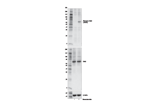
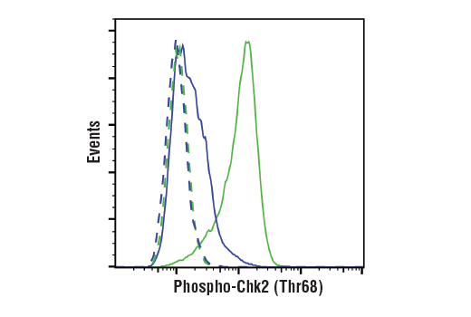
| Phospho-Chk2 (Thr68) (E8Q1A) Rabbit mAb 82263 | 20 µl |
|
H | 62 | Rabbit IgG |
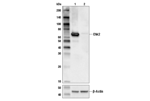
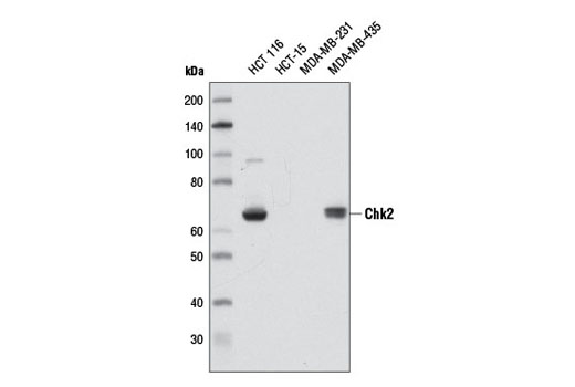
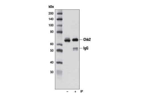
| Chk2 (D9C6) Rabbit mAb 6334 | 20 µl |
|
H | 62 | Rabbit IgG |
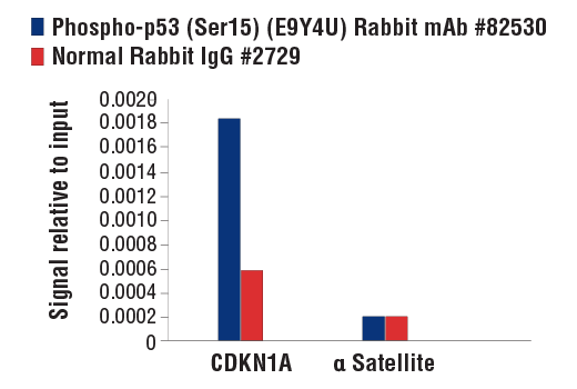
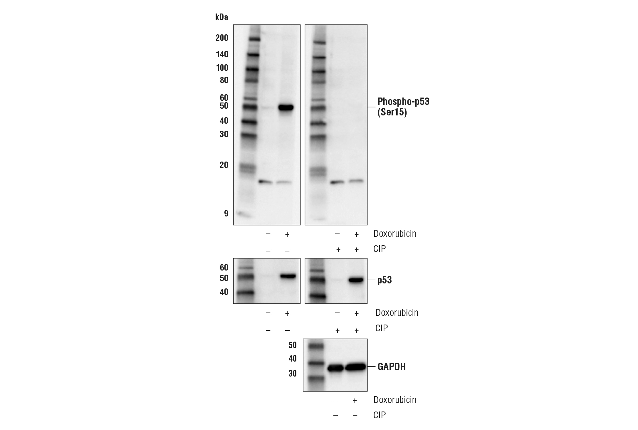
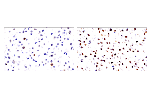
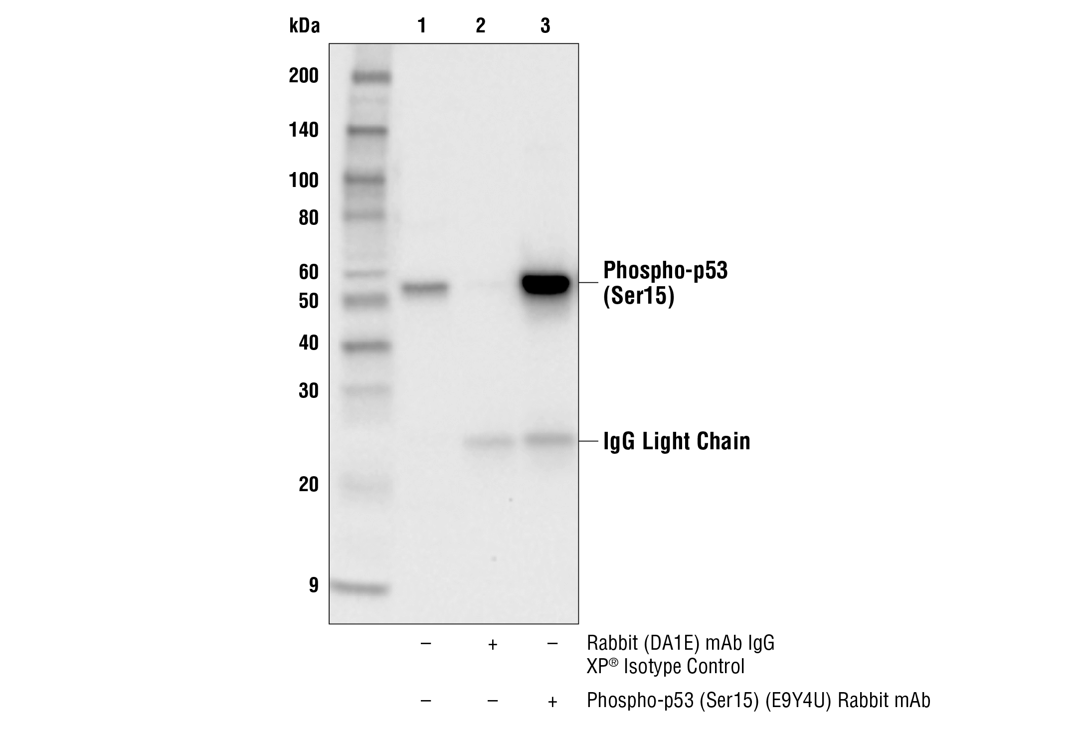
| Phospho-p53 (Ser15) (E9Y4U) Rabbit mAb 82530 | 20 µl |
|
H | 53 | Rabbit IgG |
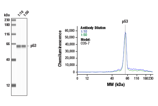
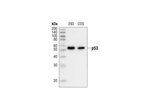
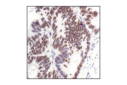
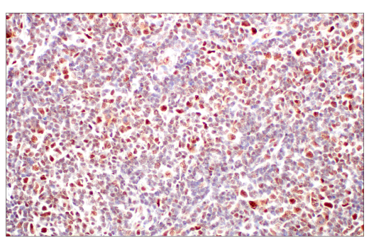
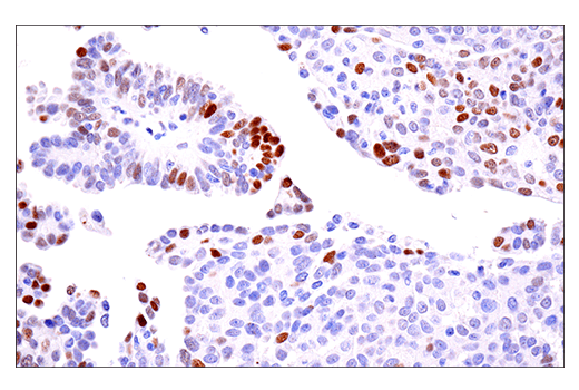
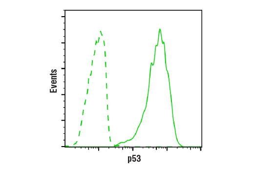
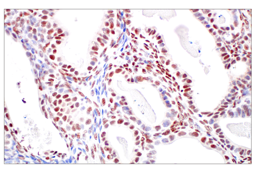
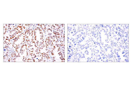
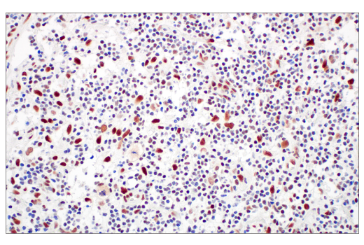
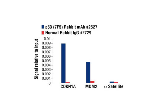
| p53 (7F5) Rabbit mAb 2527 | 20 µl |
|
H Mk | 53 | Rabbit IgG |
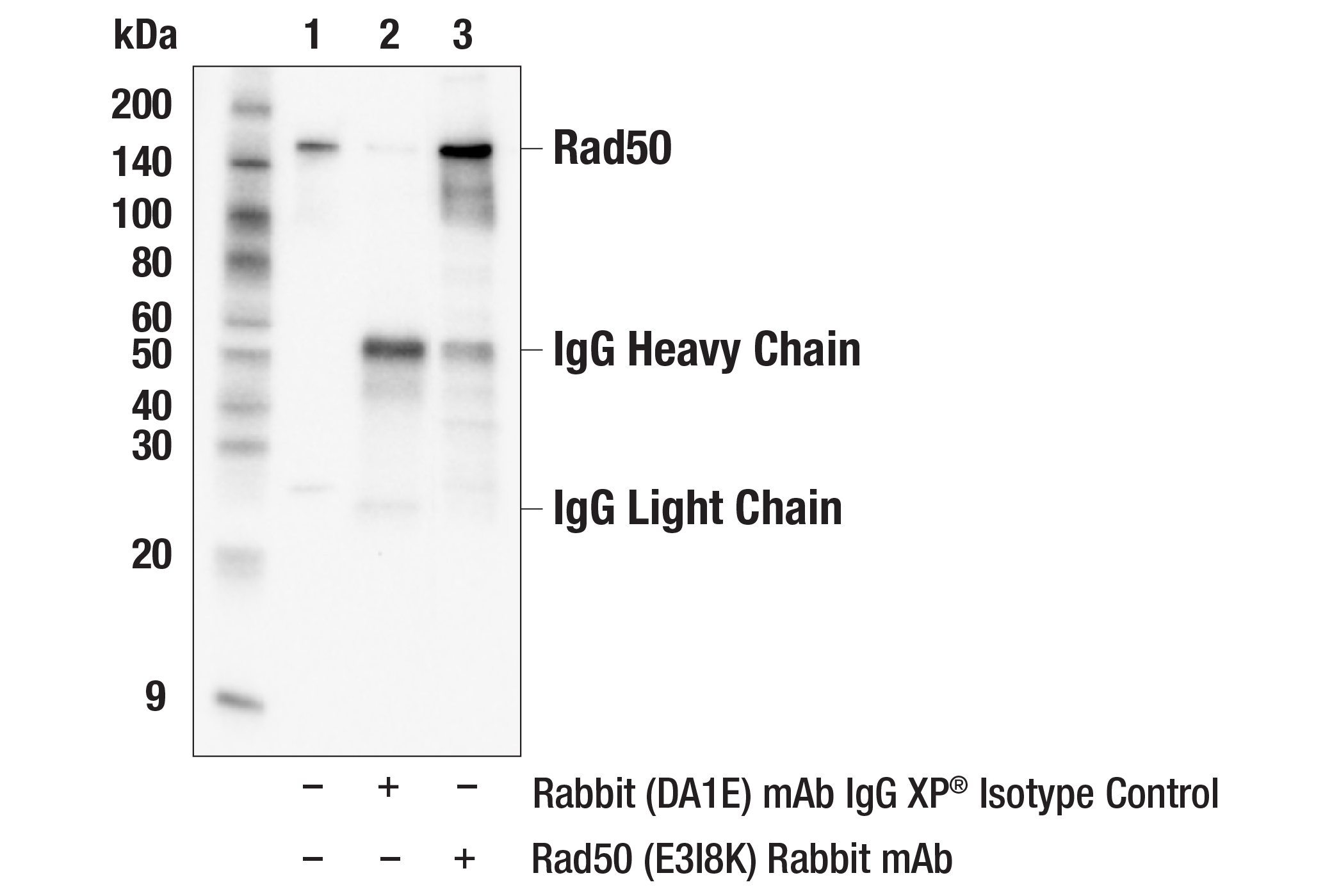
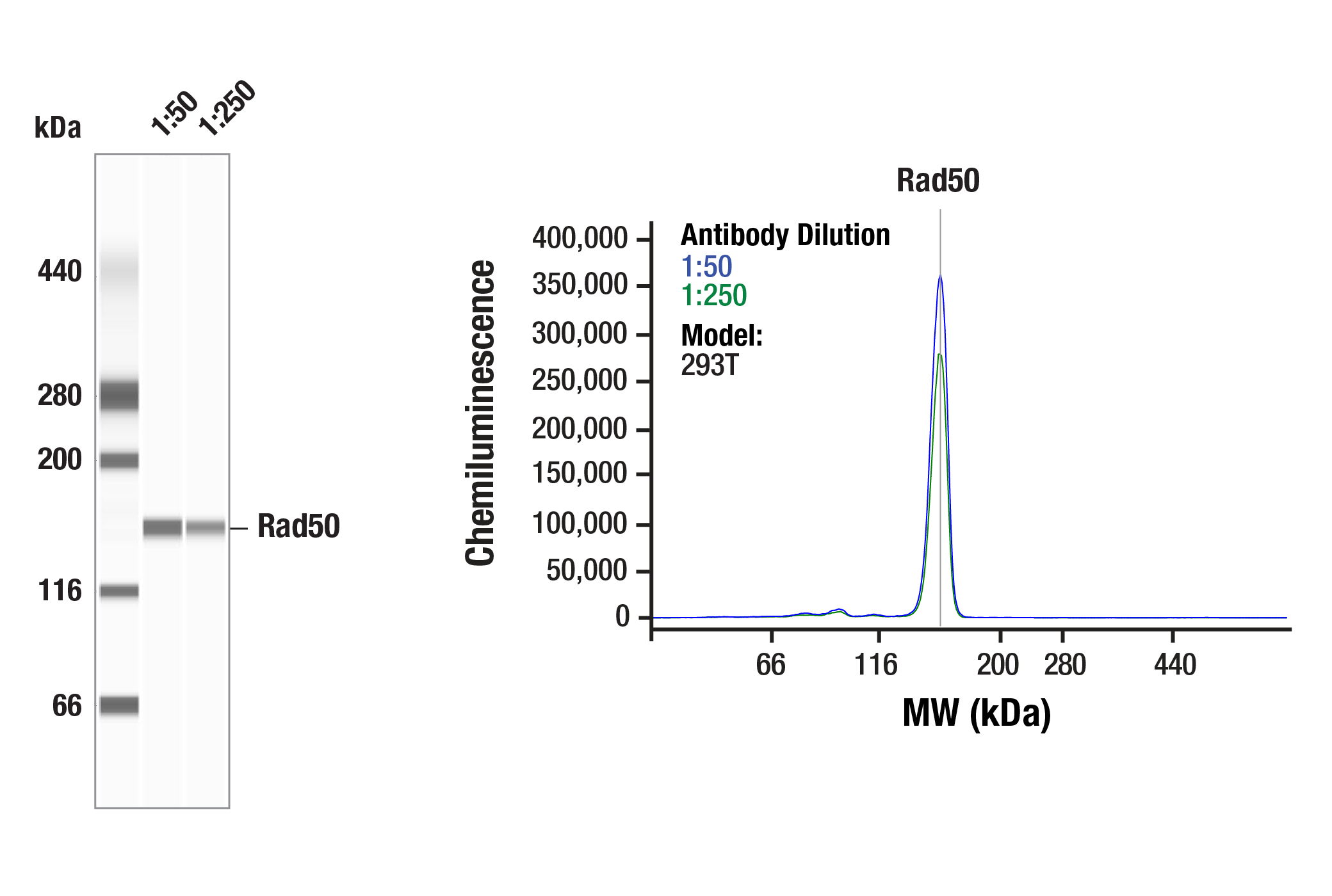
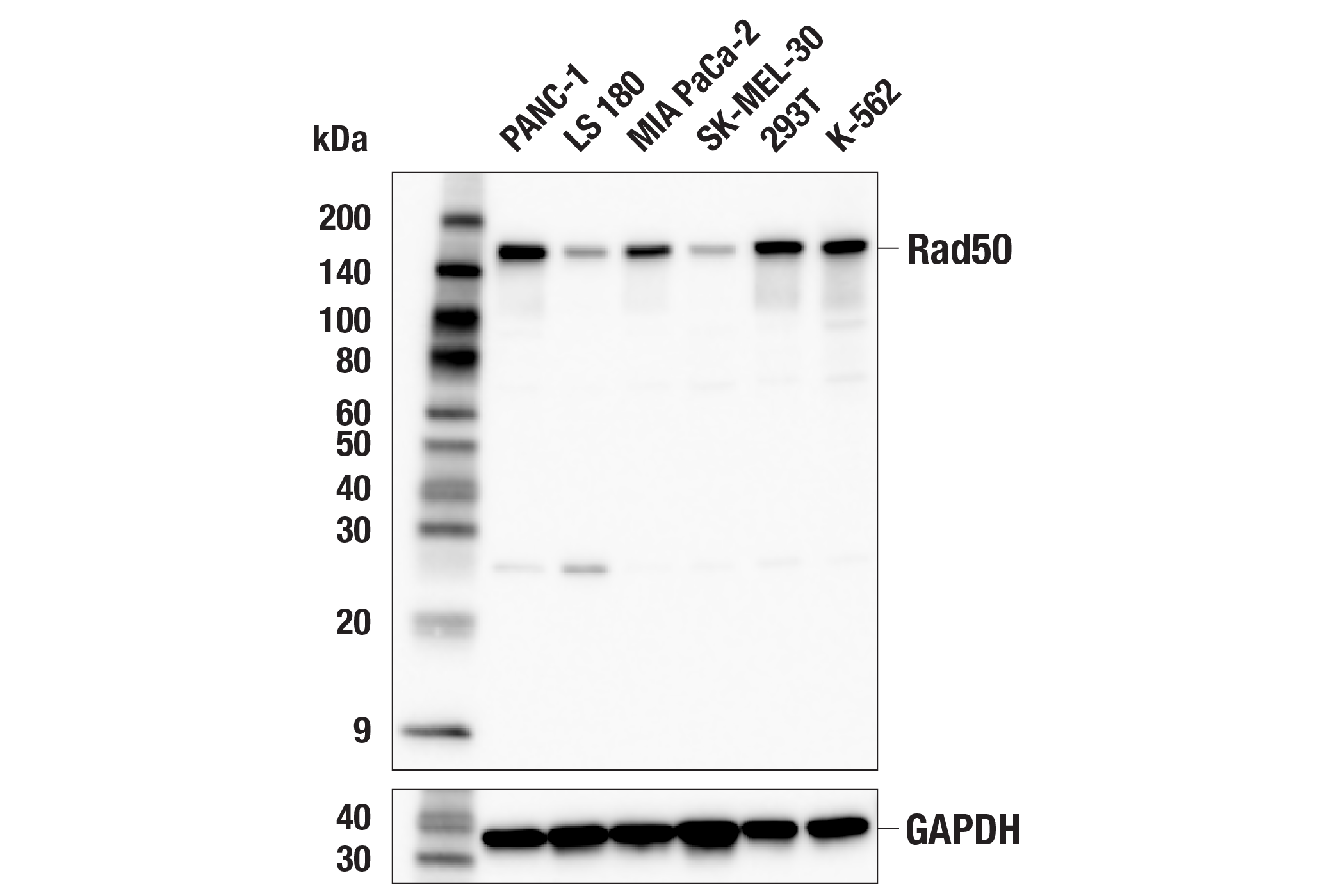
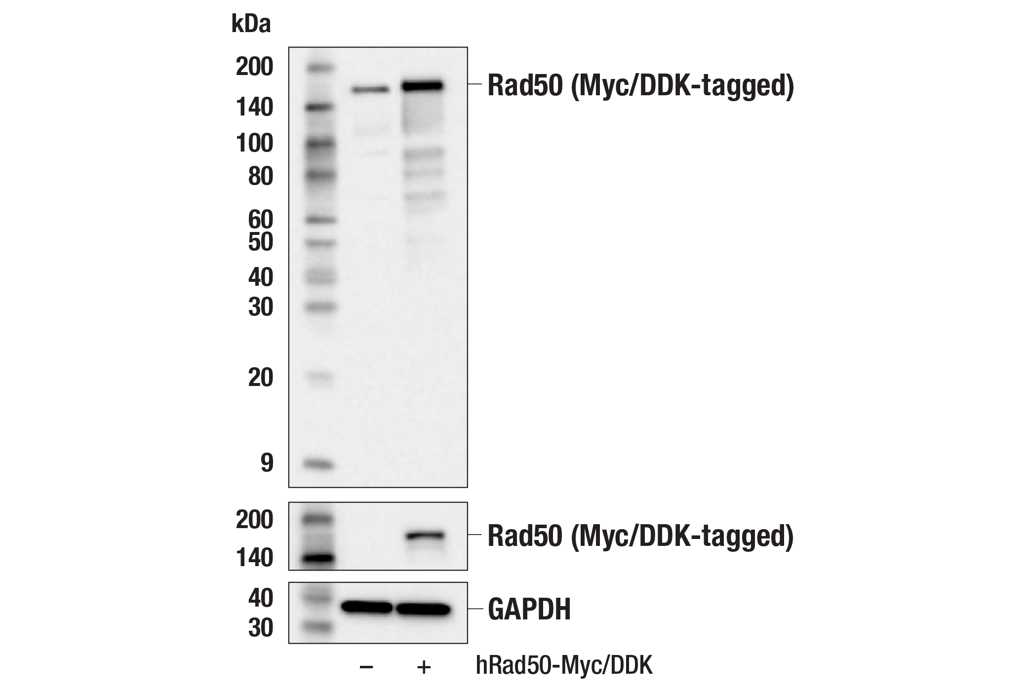
| Rad50 (E3I8K) Rabbit mAb 86225 | 20 µl |
|
H | 153 | Rabbit IgG |
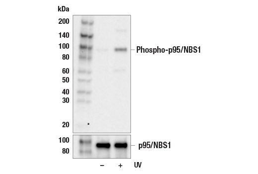
| Phospho-p95/NBS1 (Ser343) Antibody 3001 | 20 µl |
|
H | 95 | Rabbit |
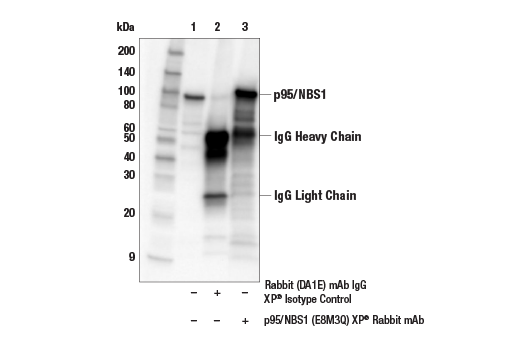
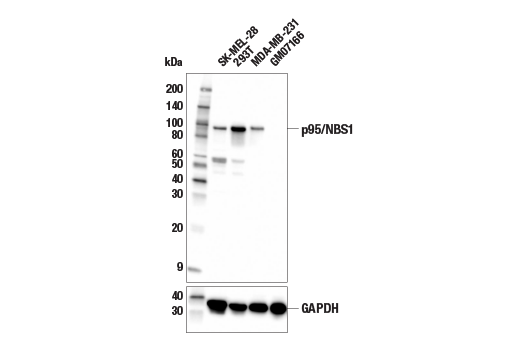
| p95/NBS1 (E8M3Q) XP® Rabbit mAb 81234 | 20 µl |
|
H | 95 | Rabbit IgG |
| Anti-rabbit IgG, HRP-linked Antibody 7074 | 100 µl |
|
Rab | Goat | |
| Phospho-Rad50 (Ser635) (F5H8B) Rabbit mAb 99215 | 20 µl |
|
H R | 153 | Rabbit IgG |
Product Information
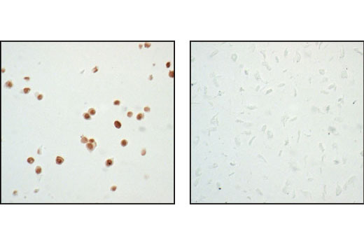
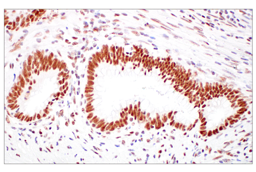
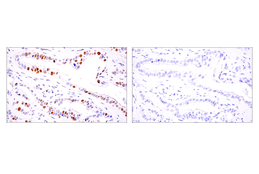
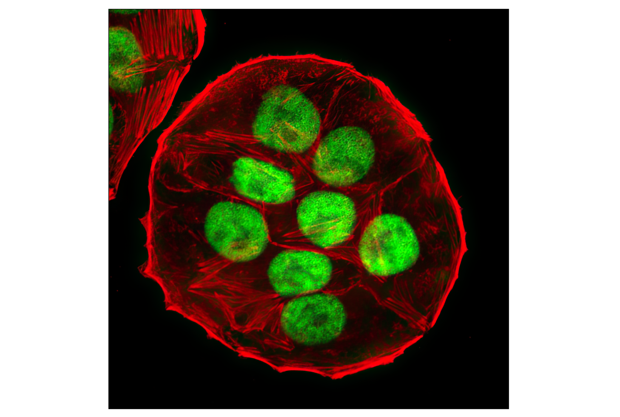
ATM (ataxia telangiectasia mutated kinase) is a serine/threonine protein kinase best known for its role in DNA repair signaling in response to DNA double-strand breaks (DSBs). When DSBs occur, the MRE11:RAD50:NBS1 (MRN) sensor complex recruits ATM to sites of DNA damage. ATM then signals to numerous effector proteins, leading to cellular responses including regulation of DNA repair, cell cycle progression, apoptosis, senescence, gene transcription. Along with ATR, DNA-PKcs, SMG1 and mTOR, ATM is a member of the PI3K-like protein kinase (PIKK) family. PIKK family members typically function in response to various types of cellular stress. Substrates of ATM are numerous, and include CHK2, AKT, p53, BRCA1 and DNA-PK (reviewed in 1,3). Inactive ATM exists as a homodimer. In response to DSBs, ATM undergoes autophosphorylation in trans at Ser1981, which leads to dissociation of the complex to become an active monomer (2). Functional DNA repair pathways are important in cellular homeostasis, and defects in these pathways cause genomic instability, which can lead to tumorigenesis (3). Inactivation of ATM results in ataxia telangiectasia (AT), a neurodegenerative disease characterized by predisposition to cancer (4).
Explore pathways related to this product.
STRING - Known and Predicted Protein-Protein Interactions.
Except as otherwise expressly agreed in a writing signed by a legally authorized representative of CST, the following terms apply to Products provided by CST, its affiliates or its distributors. Any Customer's terms and conditions that are in addition to, or different from, those contained herein, unless separately accepted in writing by a legally authorized representative of CST, are rejected and are of no force or effect.
Products are labeled with For Research Use Only or a similar labeling statement and have not been approved, cleared, or licensed by the FDA or other regulatory foreign or domestic entity, for any purpose. Customer shall not use any Product for any diagnostic or therapeutic purpose, or otherwise in any manner that conflicts with its labeling statement. Products sold or licensed by CST are provided for Customer as the end-user and solely for research and development uses. Any use of Product for diagnostic, prophylactic or therapeutic purposes, or any purchase of Product for resale (alone or as a component) or other commercial purpose, requires a separate license from CST. Customer shall (a) not sell, license, loan, donate or otherwise transfer or make available any Product to any third party, whether alone or in combination with other materials, or use the Products to manufacture any commercial products, (b) not copy, modify, reverse engineer, decompile, disassemble or otherwise attempt to discover the underlying structure or technology of the Products, or use the Products for the purpose of developing any products or services that would compete with CST products or services, (c) not alter or remove from the Products any trademarks, trade names, logos, patent or copyright notices or markings, (d) use the Products solely in accordance with CST Product Terms of Sale and any applicable documentation, and (e) comply with any license, terms of service or similar agreement with respect to any third party products or services used by Customer in connection with the Products.