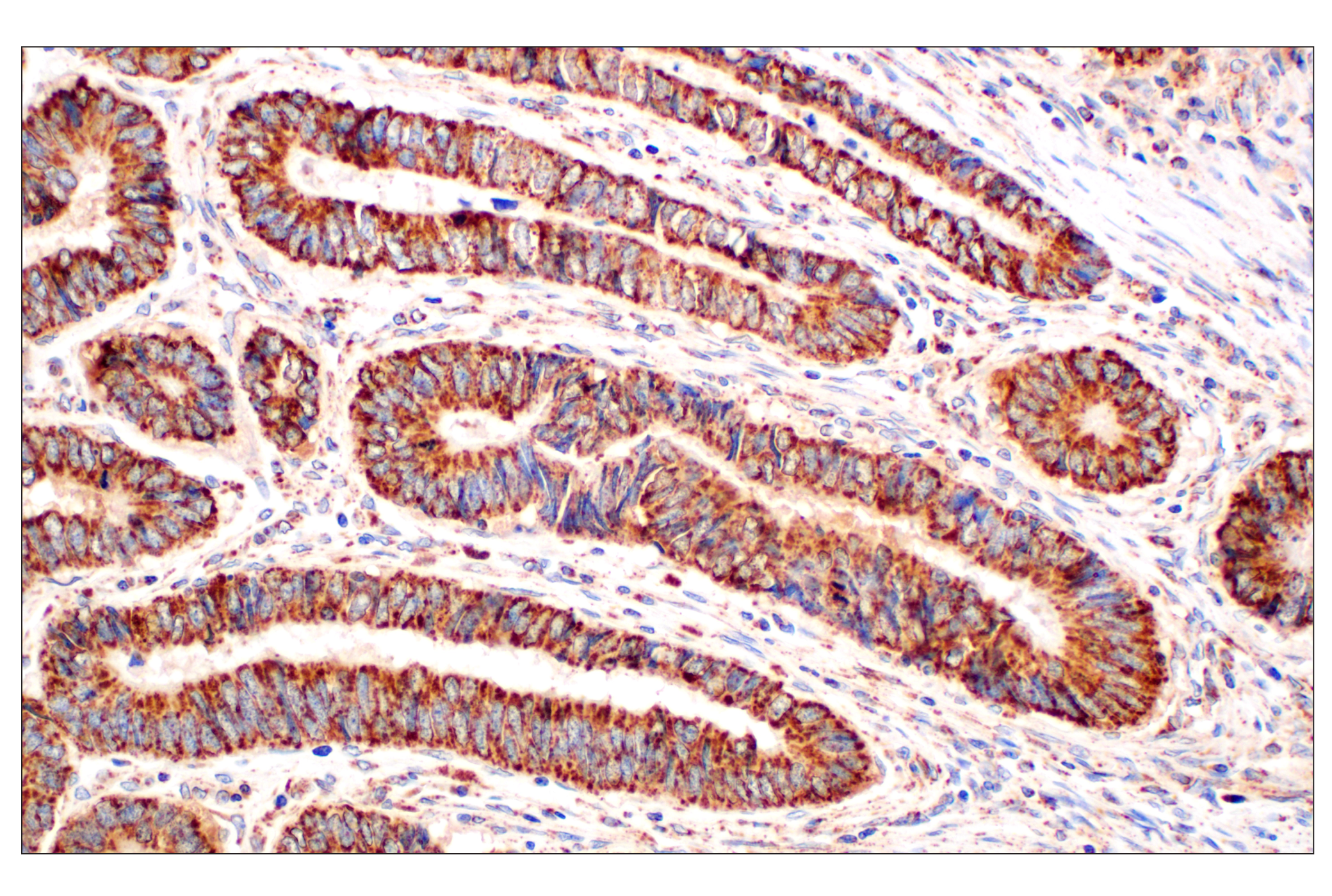
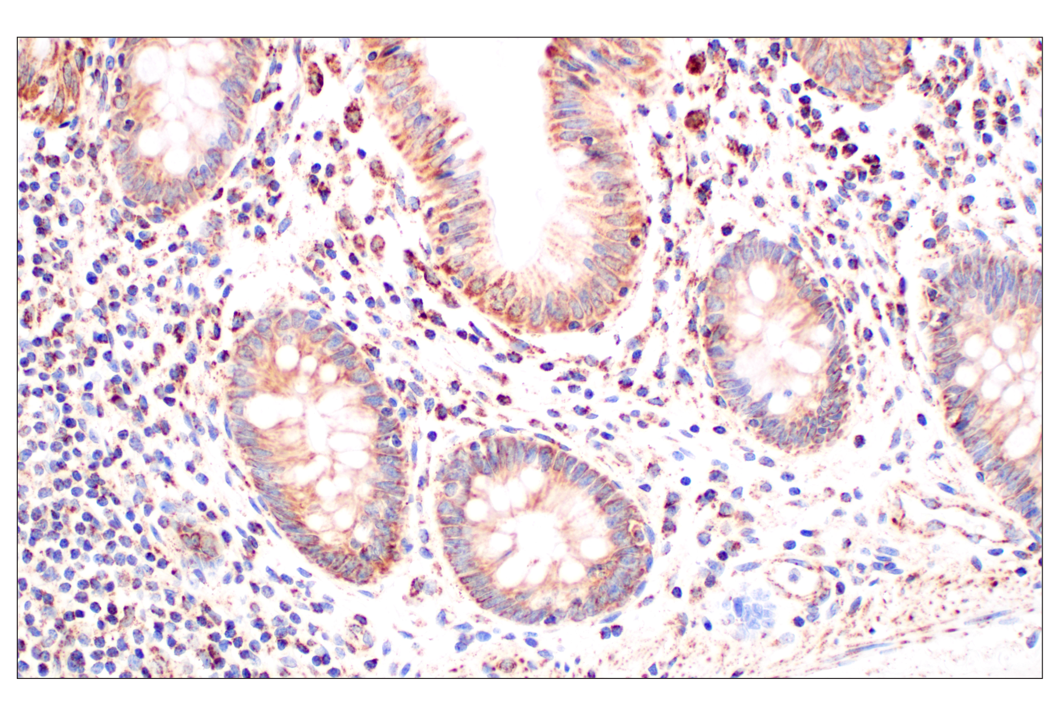
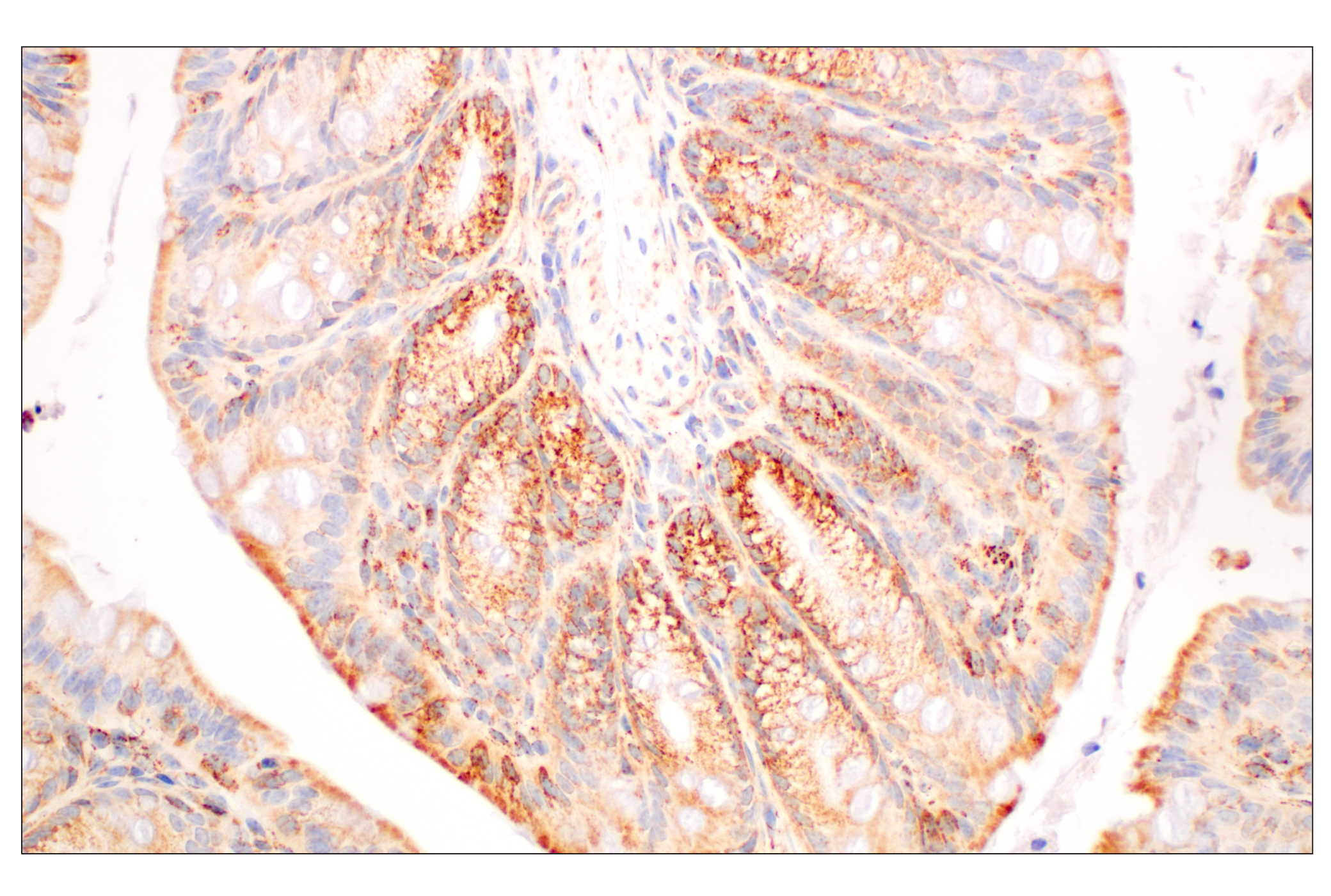
Immunohistochemical analysis of paraffin-embedded human colon adenocarcinoma using GOT2 (F4P3R) Rabbit mAb.
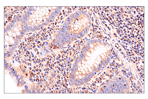
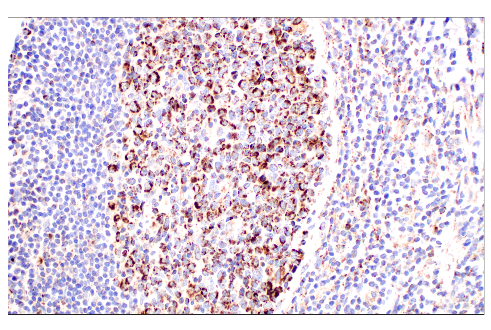
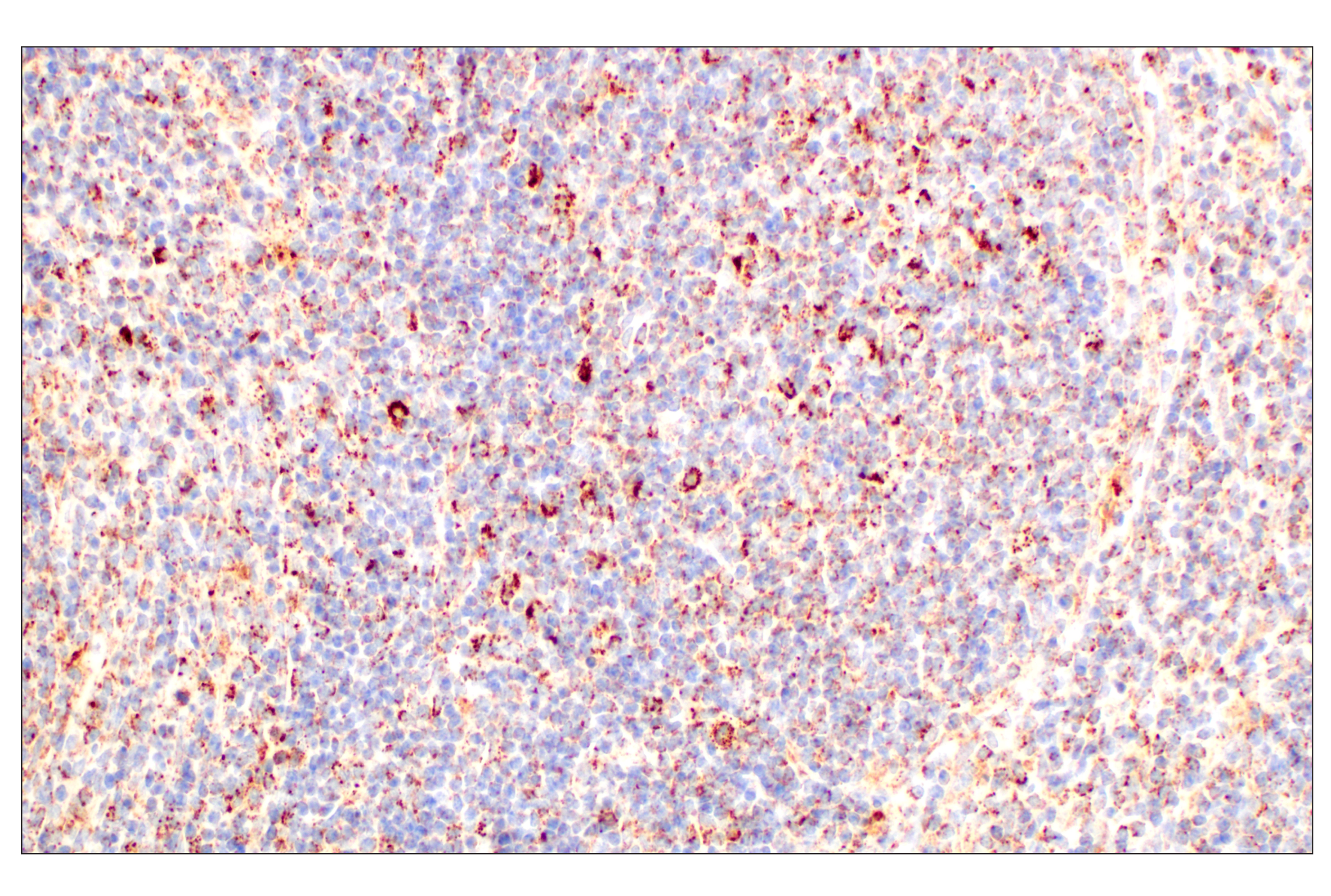
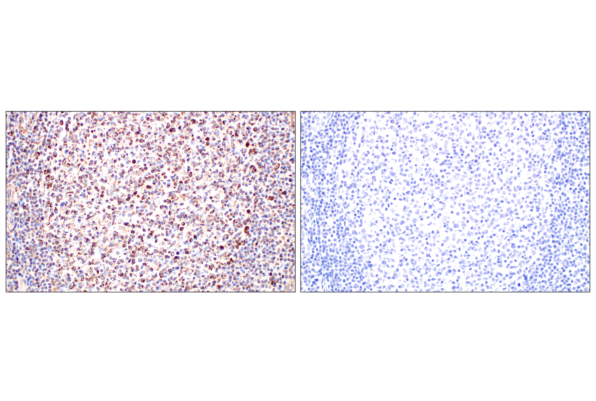
Immunohistochemical analysis of paraffin-embedded human B-cell non-Hodgkin lymphoma using GOT2 (F4P3R) Rabbit mAb.
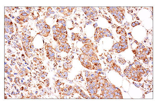
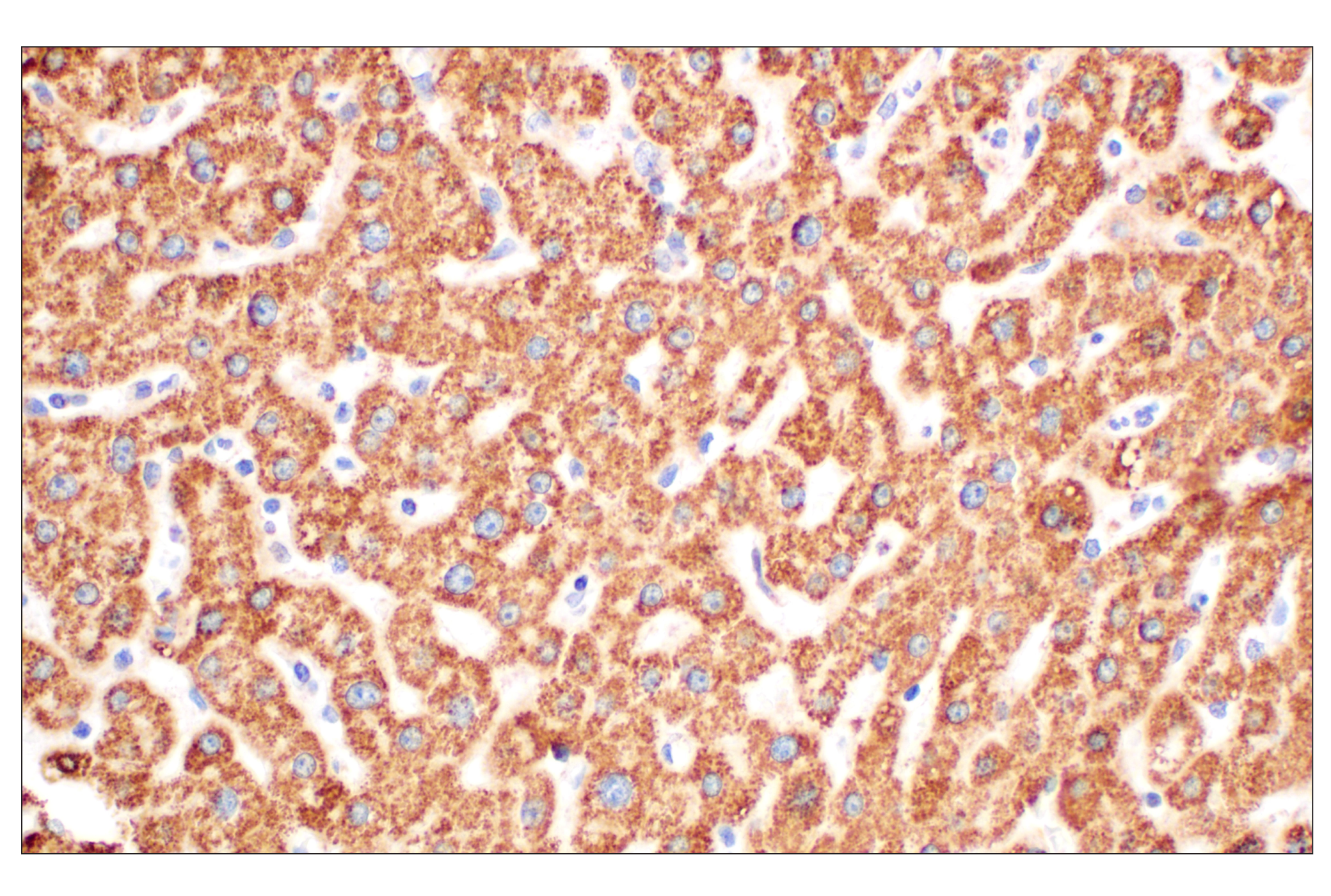
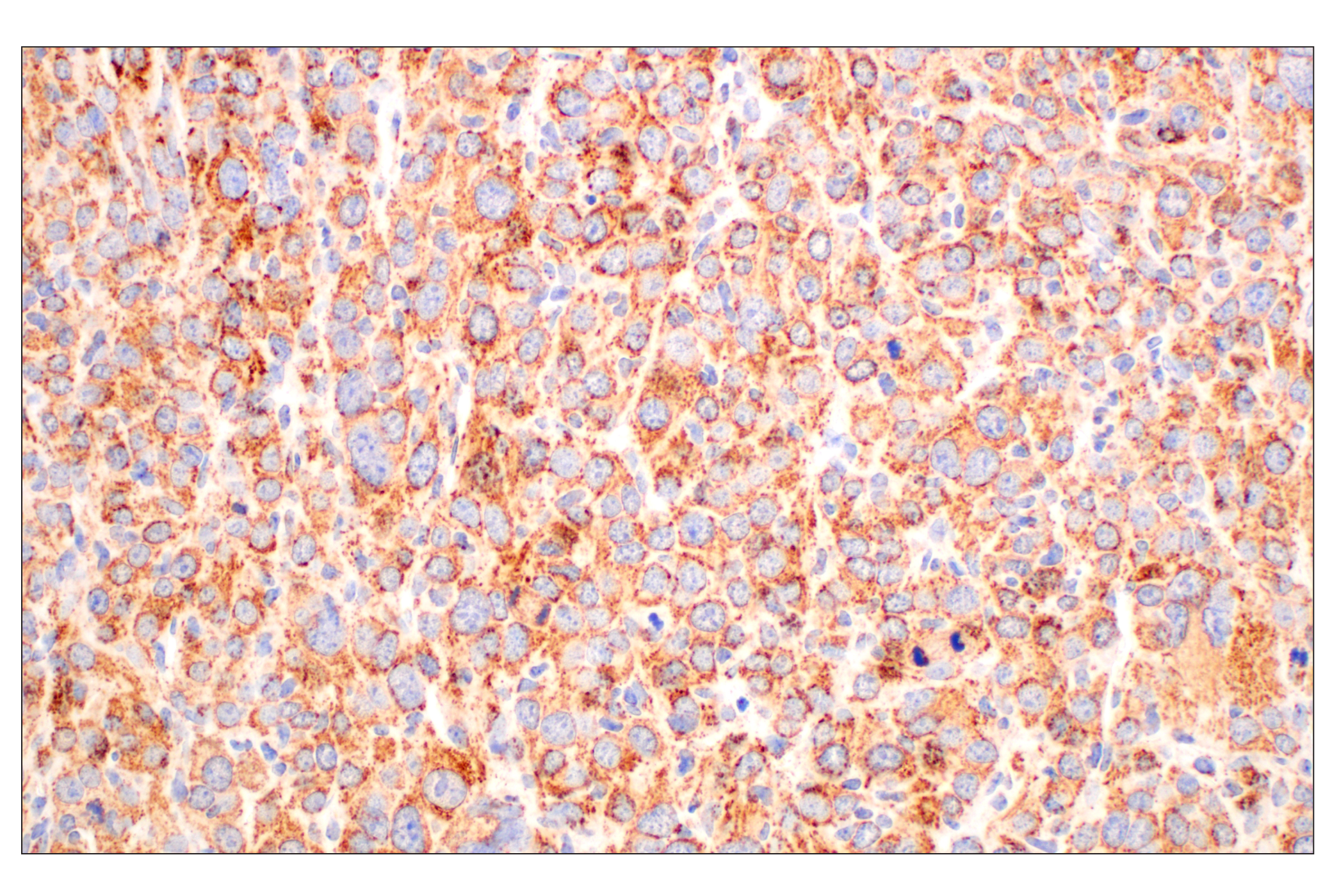
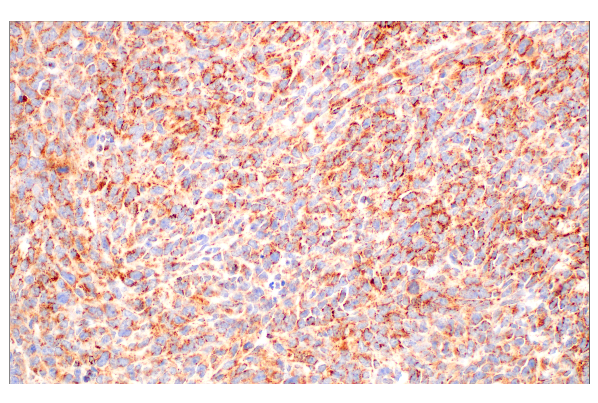
Immunohistochemical analysis of paraffin-embedded normal human adrenal gland using GOT2 (F4P3R) Rabbit mAb.
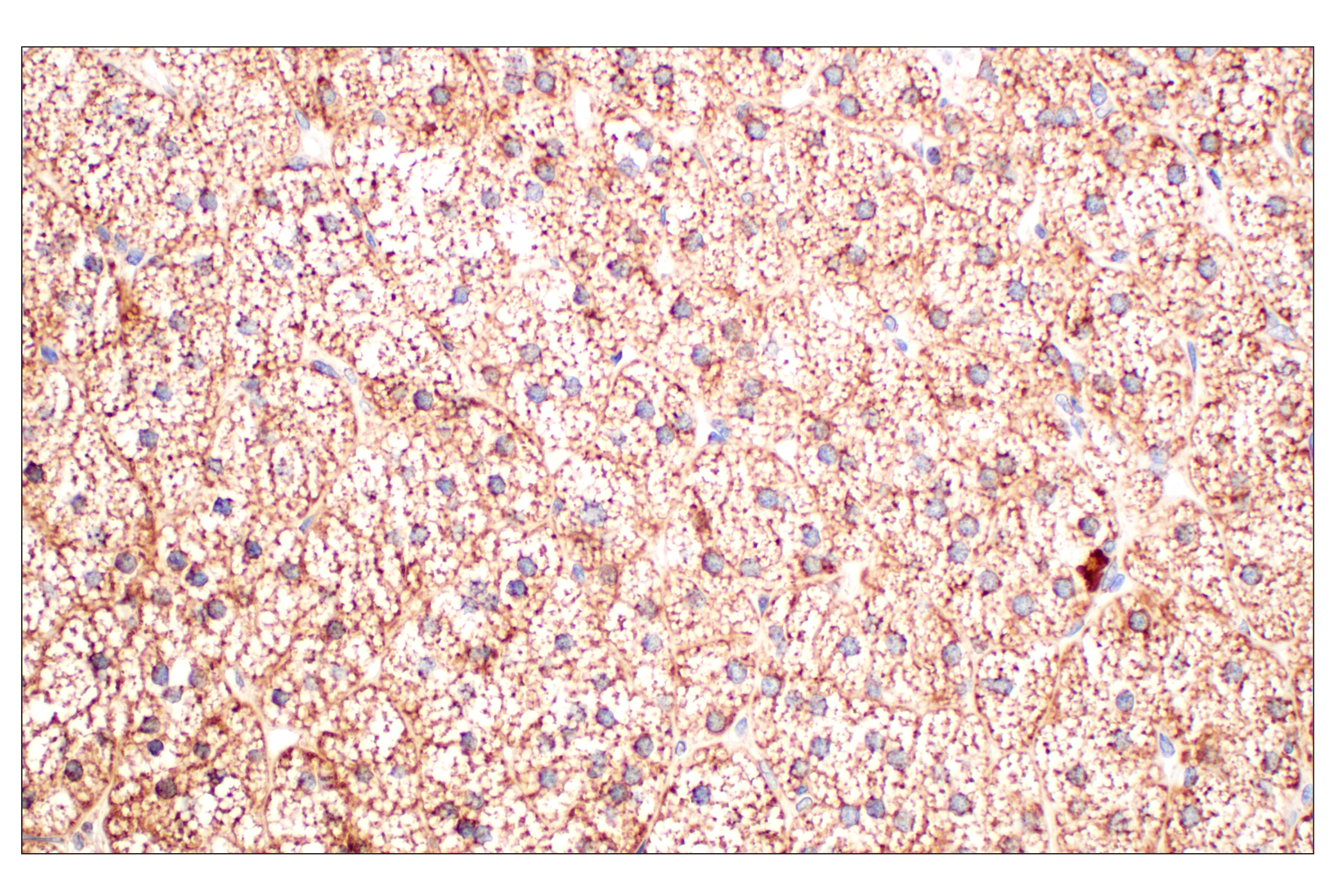
Immunohistochemical analysis of paraffin-embedded normal human epididymis using GPX4 (E5Y8K) Rabbit mAb.
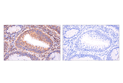
Immunohistochemical analysis of paraffin-embedded normal human stomach using GPX4 (E5Y8K) Rabbit mAb (left) compared to concentration-matched Rabbit (DA1E) mAb IgG XP® Isotype Control #3900 (right).
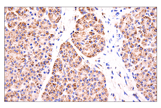
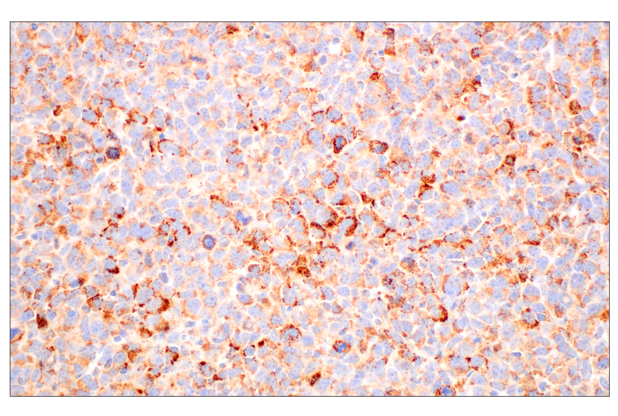
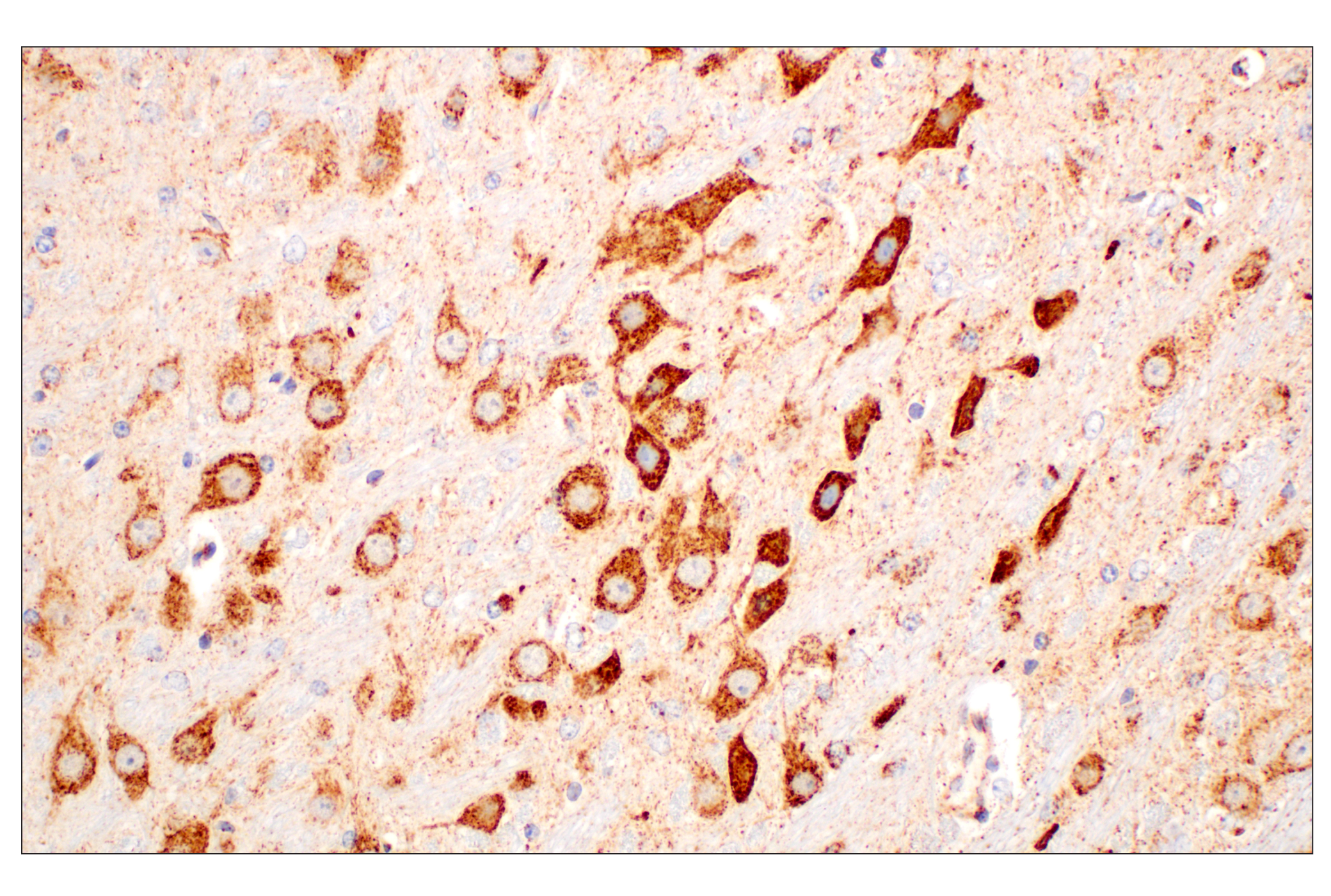
Immunohistochemical analysis of paraffin-embedded 4T1 syngeneic mammary tumor using GOT2 (F4P3R) Rabbit mAb.
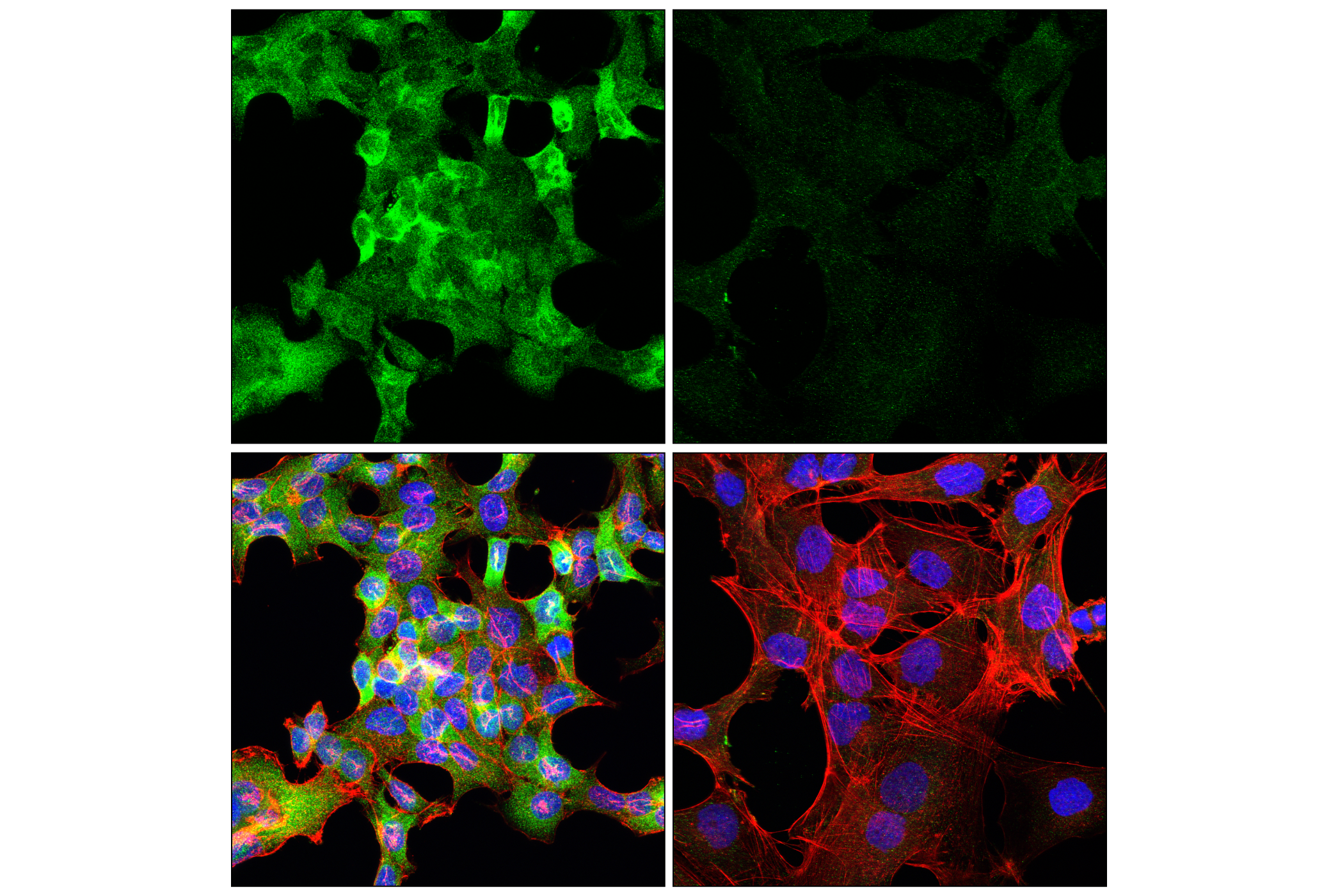
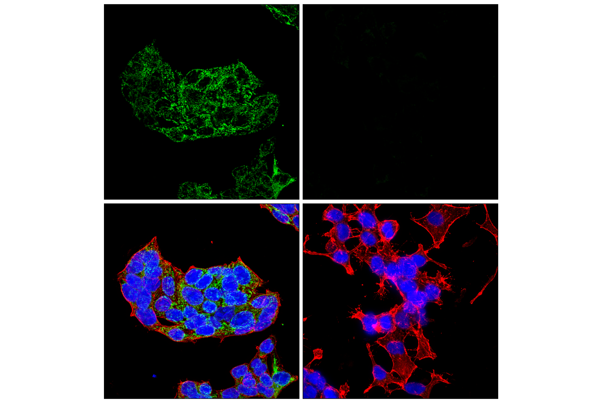
Immunohistochemical analysis of paraffin-embedded 22Rv1 cell pellet (left, high-expressing) or Daudi cell pellet (right, low-expressing) using GPX4 (E5Y8K) Rabbit mAb.
| Cat. # | Size | Qty. | Price |
|---|---|---|---|
| 84600T | 1 Kit (9 x 20 microliters) |
|
| Product Includes | Quantity | Applications | Reactivity | MW(kDa) | Isotype |
|---|---|---|---|---|---|
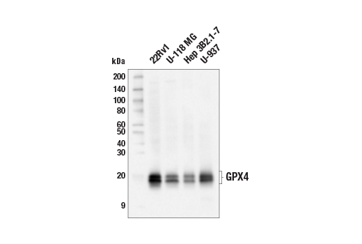
| GPX4 (E5Y8K) Rabbit mAb 59735 | 20 µl |
|
H M R | 20, 22 | Rabbit IgG |
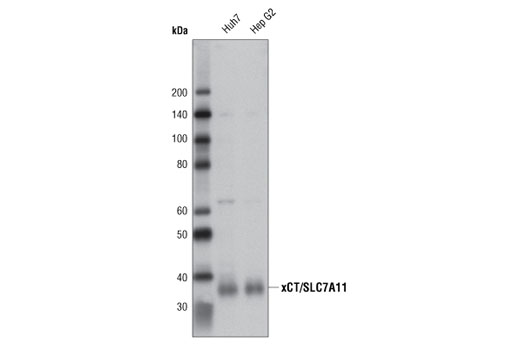
| xCT/SLC7A11 (D2M7A) Rabbit mAb 12691 | 20 µl |
|
H | 35 | Rabbit IgG |
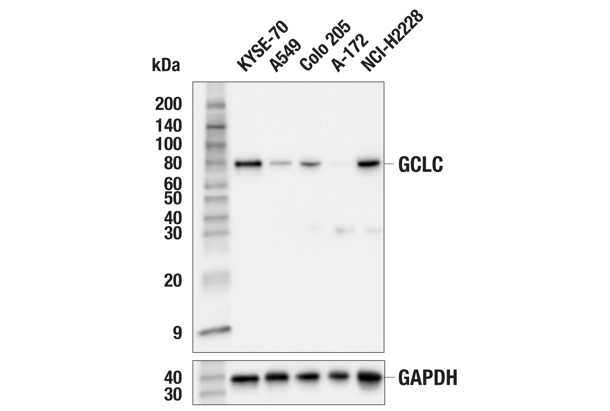
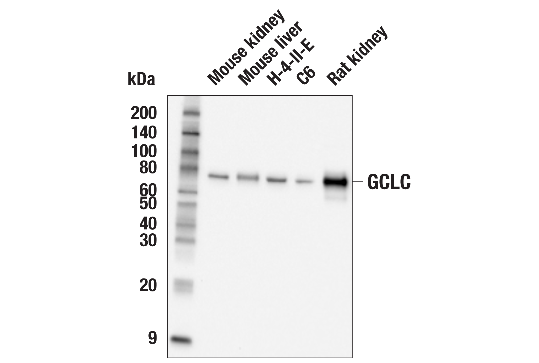
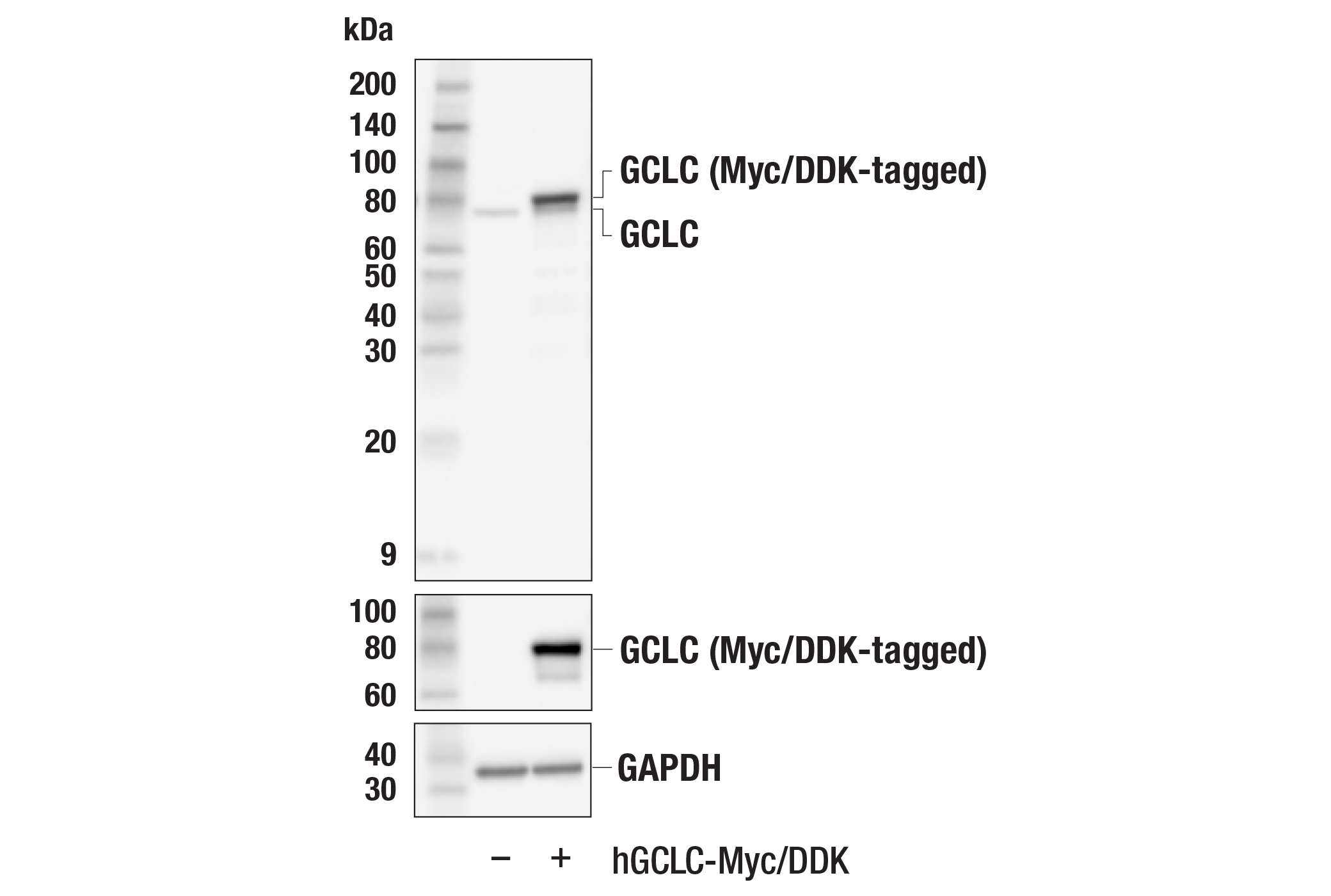
| GCLC (E2Y7D) Rabbit mAb 52183 | 20 µl |
|
H M R | 78 | Rabbit IgG |
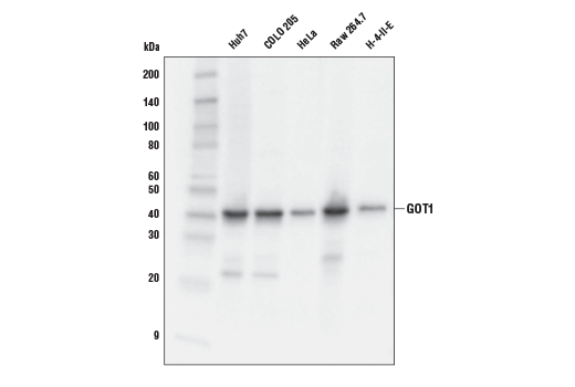
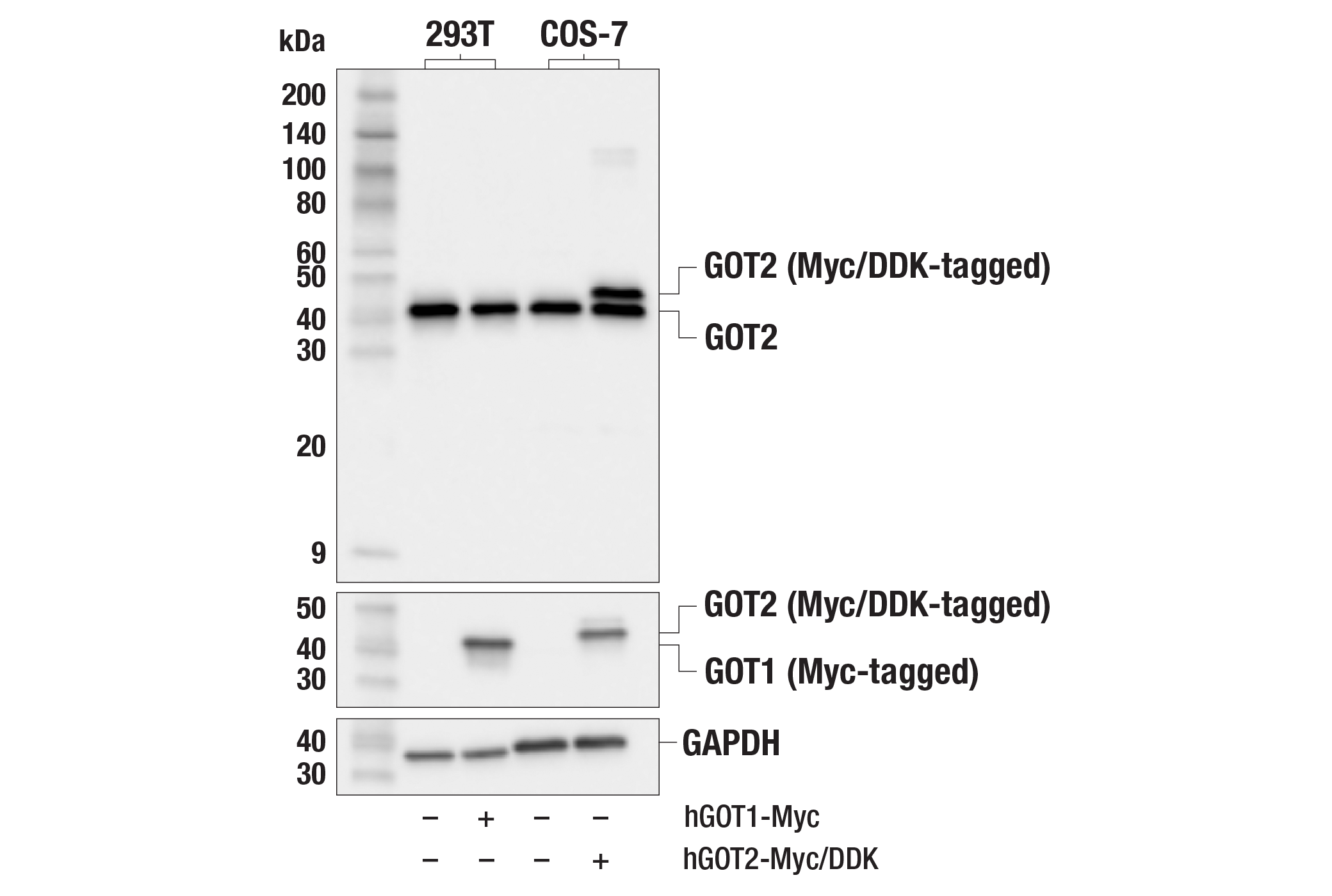
| GOT1 (E4A4O) Rabbit mAb 34423 | 20 µl |
|
H M R Mk | 41 | Rabbit IgG |
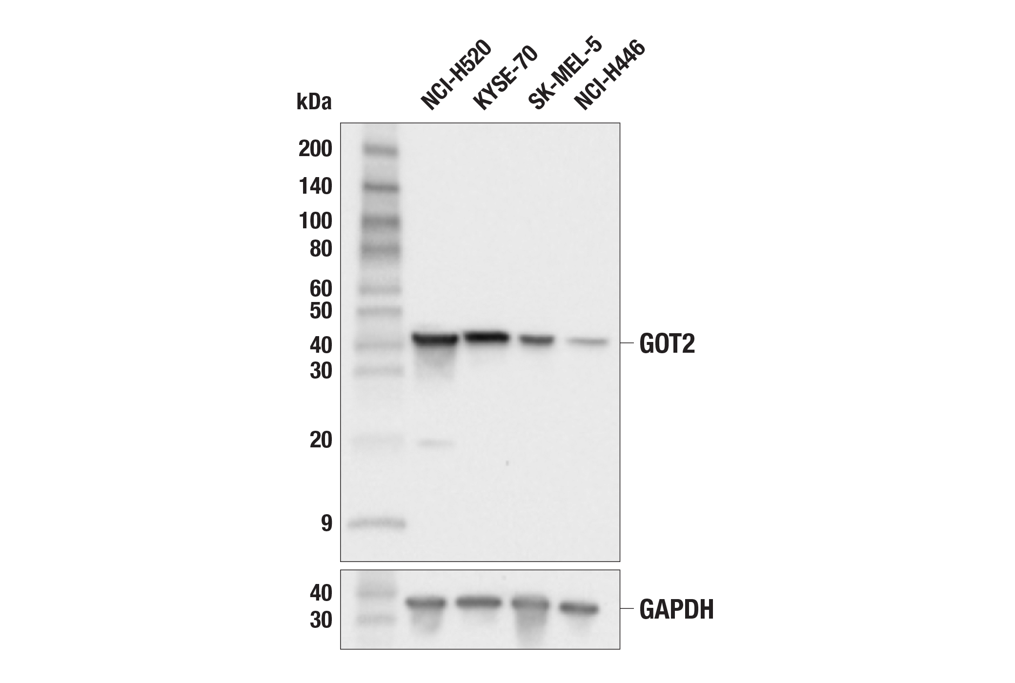
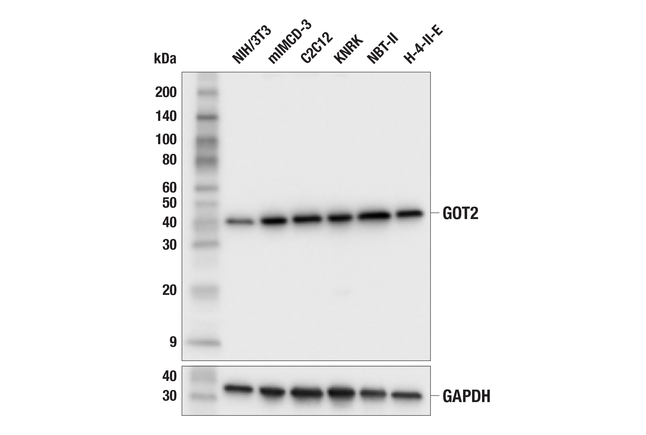
| GOT2 (F4P3R) Rabbit mAb 39627 | 20 µl |
|
H M R | 41 | Rabbit IgG |
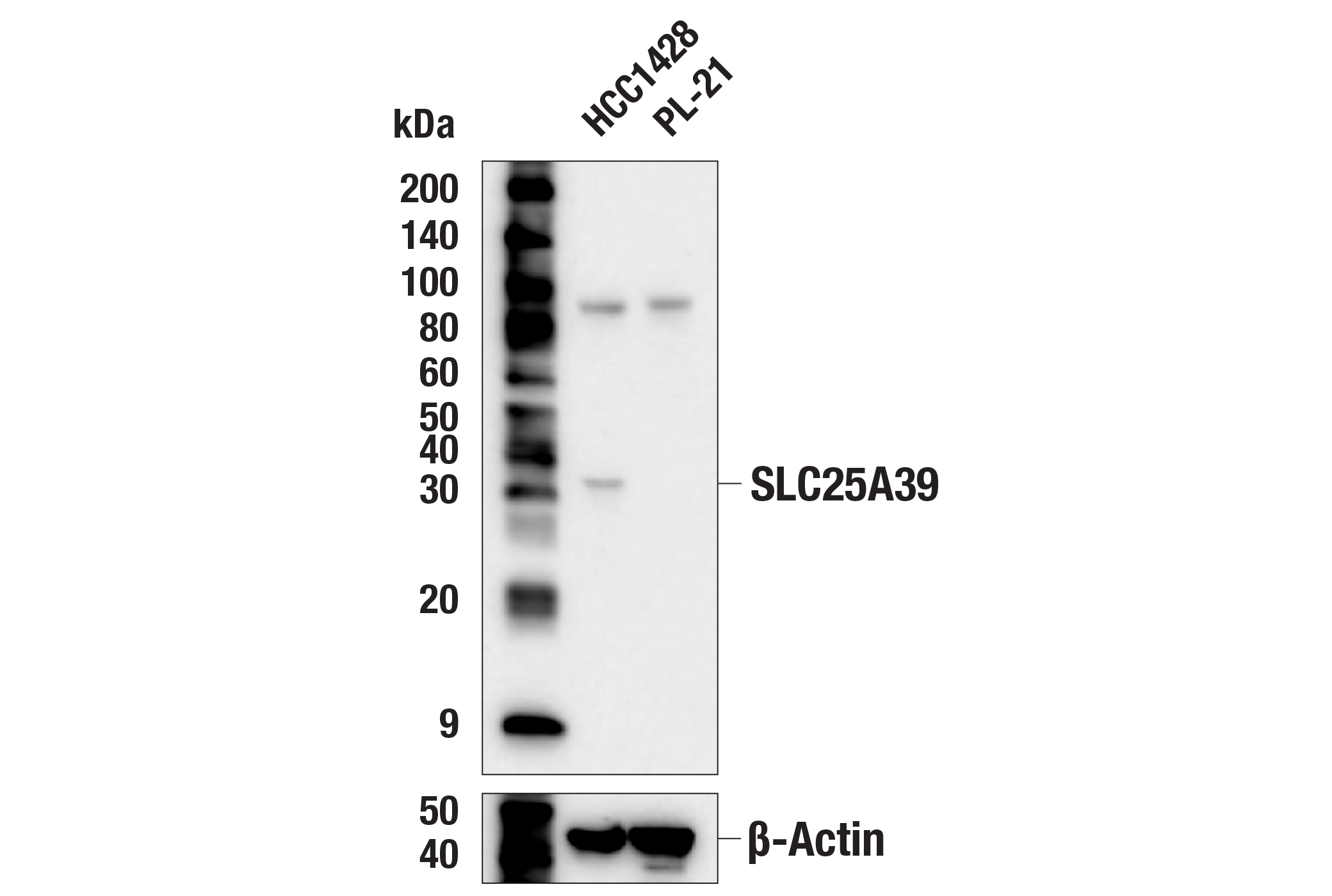
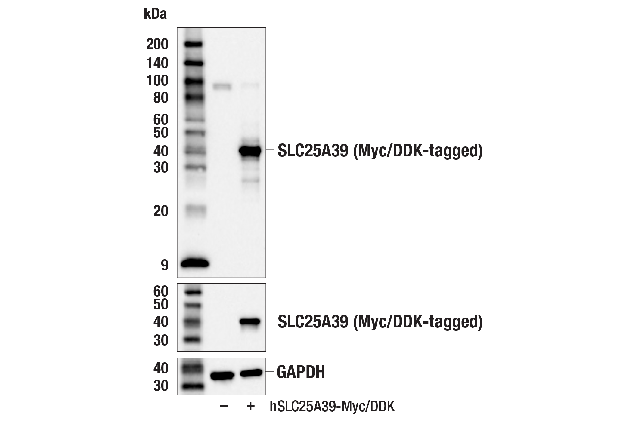
| SLC25A39 (F2F6O) Rabbit mAb 33871 | 20 µl |
|
H | 36 | Rabbit IgG |
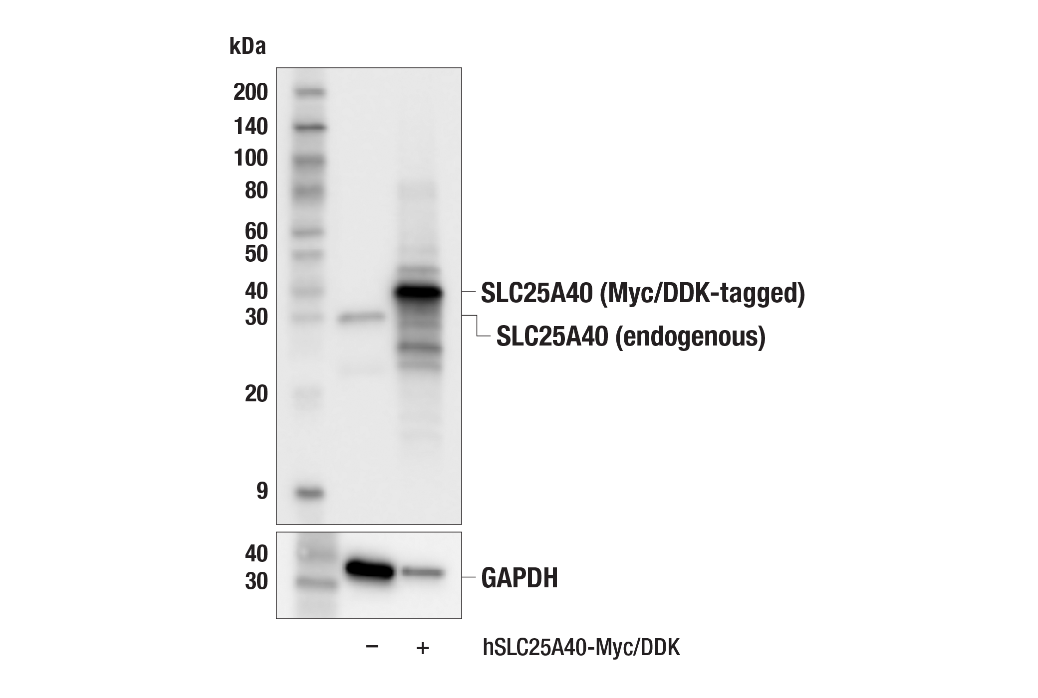
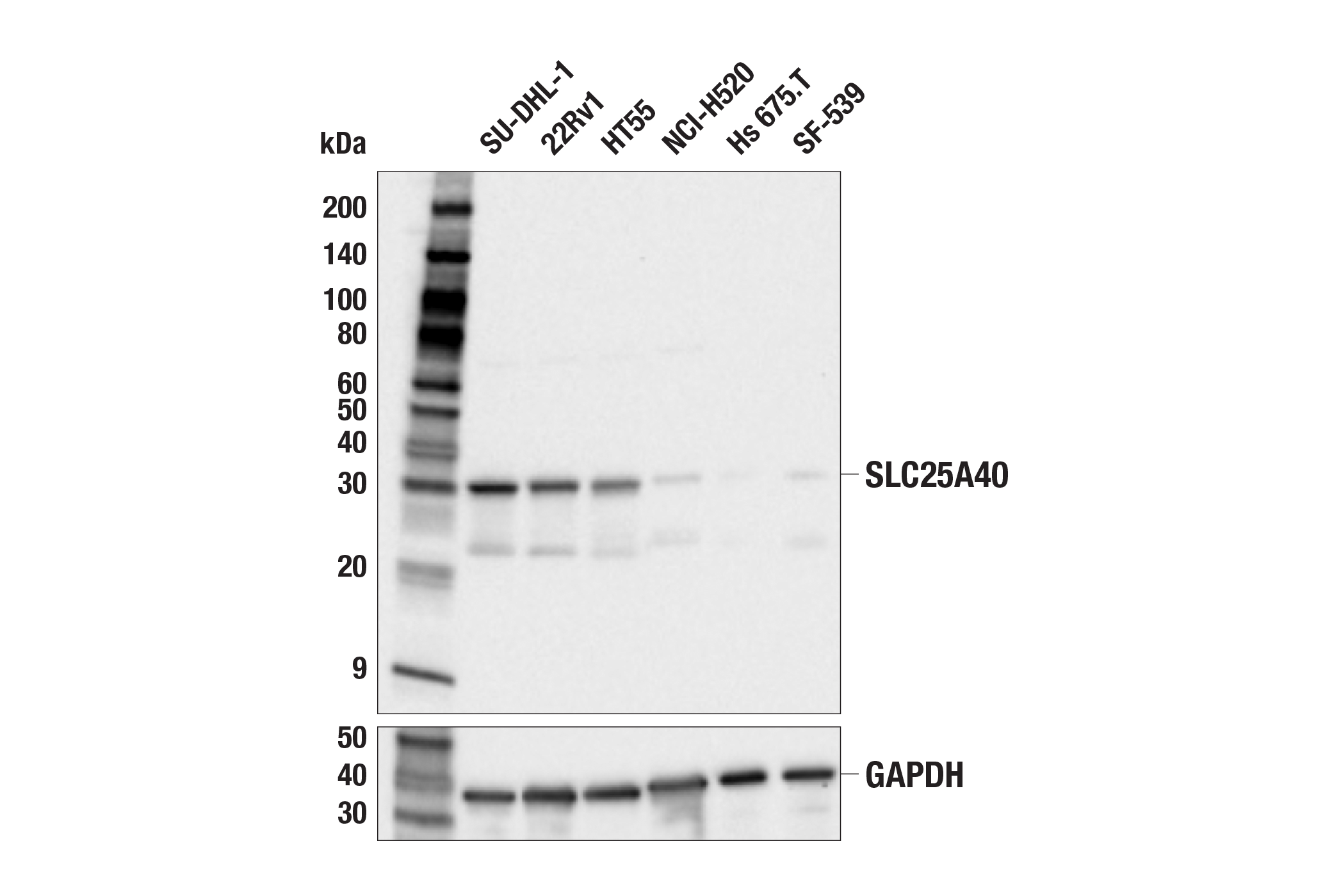
| SLC25A40 (E9C7Y) Rabbit mAb 38244 | 20 µl |
|
H | 32 | Rabbit IgG |
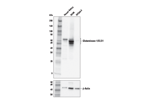
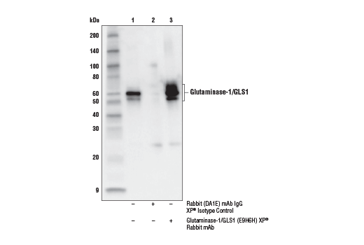
| Glutaminase-1/GLS1 (E9H6H) XP® Rabbit mAb 56750 | 20 µl |
|
H Mk | 55-65 | Rabbit IgG |
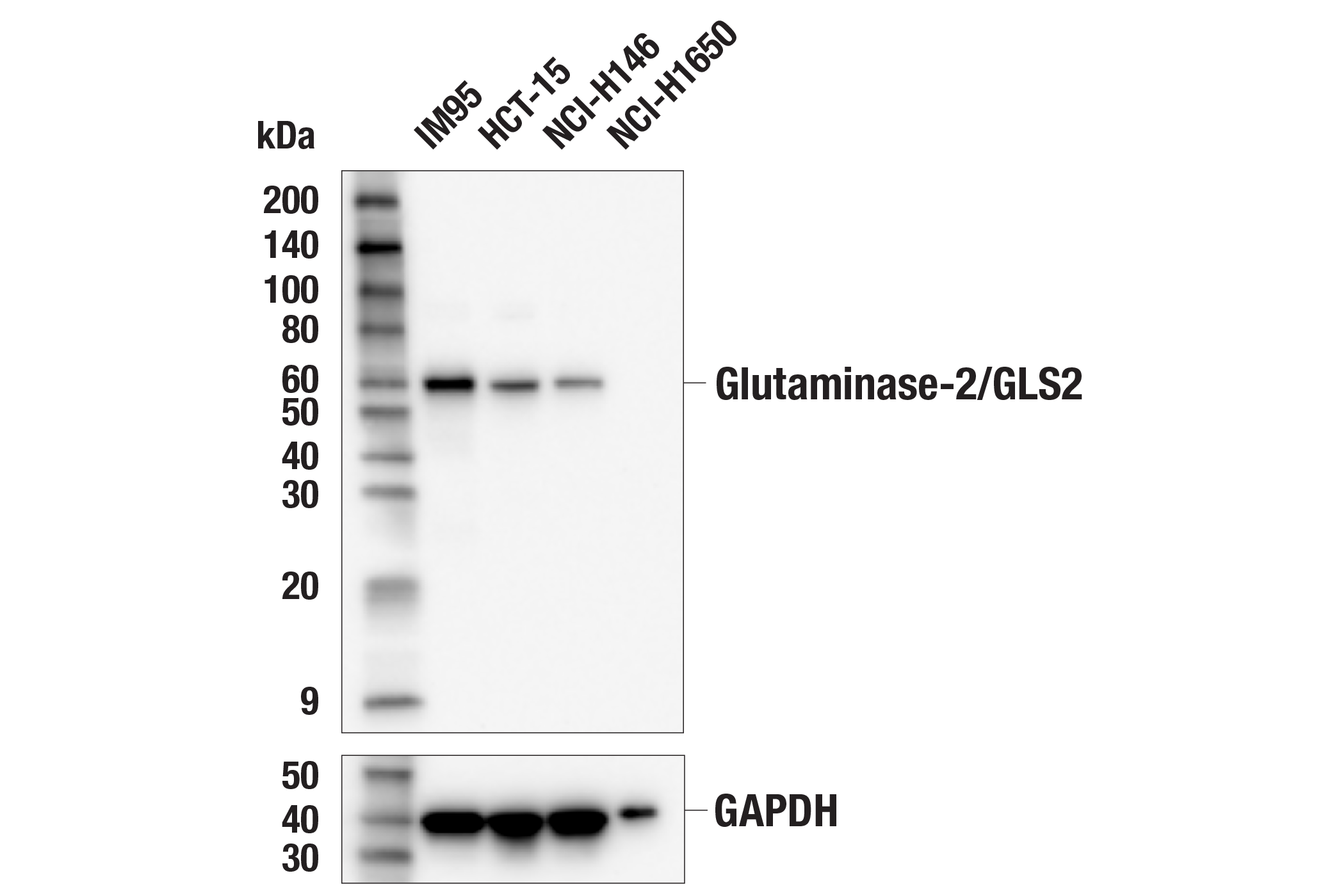
| Glutaminase-2/GLS2 (E9C7V) Rabbit mAb 85934 | 20 µl |
|
H | 60 | Rabbit IgG |
| Anti-rabbit IgG, HRP-linked Antibody 7074 | 100 µl |
|
Rab | Goat |
Product Information
Ferroptosis is an iron-dependent form of regulated cell death associated with increased lipid peroxides (reviewed in 1,2). Free divalent iron (Fe2+) can lead to spontaneous lipid peroxidation through a Fenton reaction. Ferroptosis is regulated by signaling pathways that control iron storage and oxidative stress. The glutathione peroxidase pathway has been identified as a key antioxidant defense pathway triggering ferroptosis. The compound RSL3, which directly inhibits GPX4, was identified as an activator of ferroptosis (3). GPX4 converts reduced glutathione (GSH) into oxidized glutathione (GSSH) and reduces cytotoxic lipid peroxides. The glutathione peroxidase pathway is further regulated by System Xc-, an amino acid antiporter consisting of a disulfide-linked heterodimer of xCT/SLC7A11 and SLC3A2/4F2hc/CD98, and is inhibited by the ferroptosis inducer erastin (4). Glutamate-cysteine ligase catalytic subunit (GCLC) catalyzes the ligation of glutamate and cysteine, the first and rate-limiting step in glutathione biosynthesis (5). SLC25A39 and its paralogue SLC25A40 were found to be essential for mitochondrial import of glutathione, providing protection against oxidative stress and mitochondrial dysfunction (6,7). Glutaminase catalyzes the conversion of glutamine to glutamate, the first and rate-limiting step of glutaminolysis (8). Both kidney-type glutaminase (GLS1) and liver-type glutaminase (GLS2) have been shown to act as tumor suppressors in some conditions (8). They have also been implicated in the regulation of ferroptosis (9,10). Glutamate oxaloacetate transaminase 1 and 2 (GOT1/GOT2) enzymes act downstream in glutamine metabolism in reactions producing aspartate. GOT1 catalyzes the cytoplasmic interconversion of aspartate and oxaloacetate (11). GOT1 inhibition promotes pancreatic cell death by ferroptosis (12). GOT2 catalyzes the conversion of oxaloacetate to aspartate in the mitochondria and has been shown to be an important regulator of cancer metabolism (13).
Explore pathways related to this product.
STRING - Known and Predicted Protein-Protein Interactions.
Except as otherwise expressly agreed in a writing signed by a legally authorized representative of CST, the following terms apply to Products provided by CST, its affiliates or its distributors. Any Customer's terms and conditions that are in addition to, or different from, those contained herein, unless separately accepted in writing by a legally authorized representative of CST, are rejected and are of no force or effect.
Products are labeled with For Research Use Only or a similar labeling statement and have not been approved, cleared, or licensed by the FDA or other regulatory foreign or domestic entity, for any purpose. Customer shall not use any Product for any diagnostic or therapeutic purpose, or otherwise in any manner that conflicts with its labeling statement. Products sold or licensed by CST are provided for Customer as the end-user and solely for research and development uses. Any use of Product for diagnostic, prophylactic or therapeutic purposes, or any purchase of Product for resale (alone or as a component) or other commercial purpose, requires a separate license from CST. Customer shall (a) not sell, license, loan, donate or otherwise transfer or make available any Product to any third party, whether alone or in combination with other materials, or use the Products to manufacture any commercial products, (b) not copy, modify, reverse engineer, decompile, disassemble or otherwise attempt to discover the underlying structure or technology of the Products, or use the Products for the purpose of developing any products or services that would compete with CST products or services, (c) not alter or remove from the Products any trademarks, trade names, logos, patent or copyright notices or markings, (d) use the Products solely in accordance with CST Product Terms of Sale and any applicable documentation, and (e) comply with any license, terms of service or similar agreement with respect to any third party products or services used by Customer in connection with the Products.