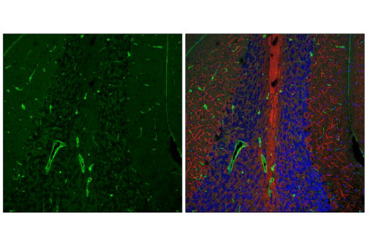
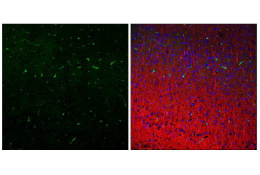
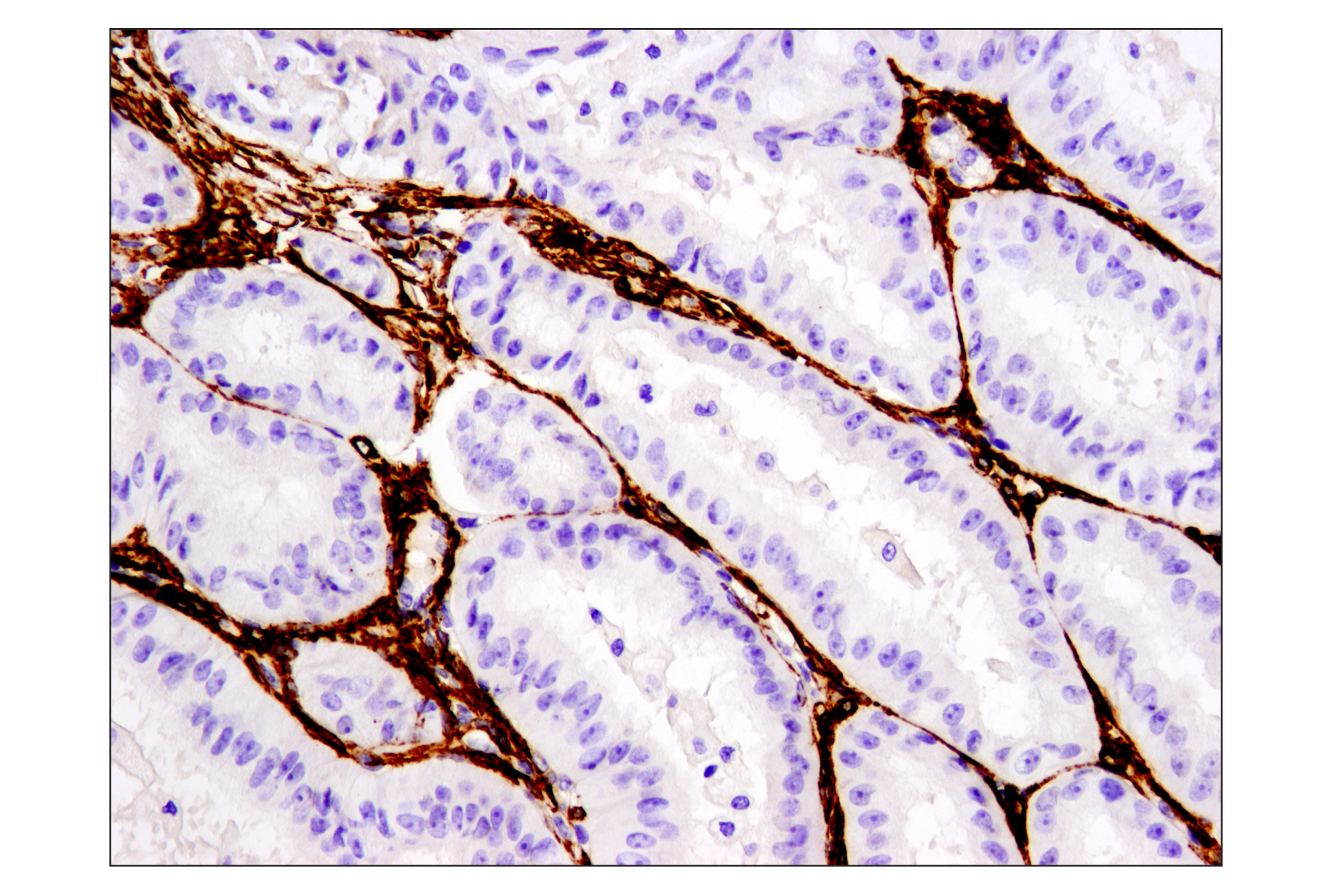
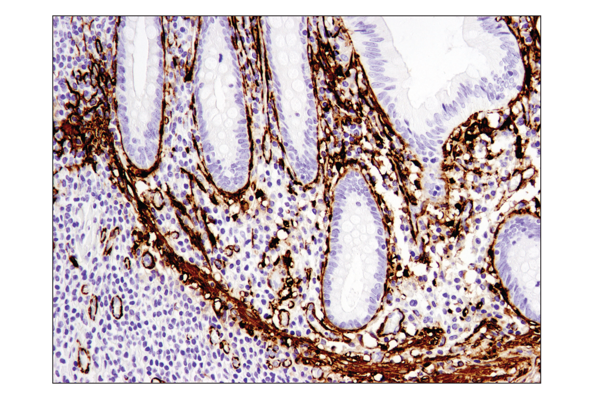
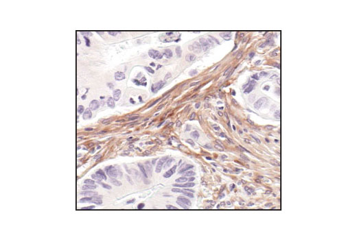
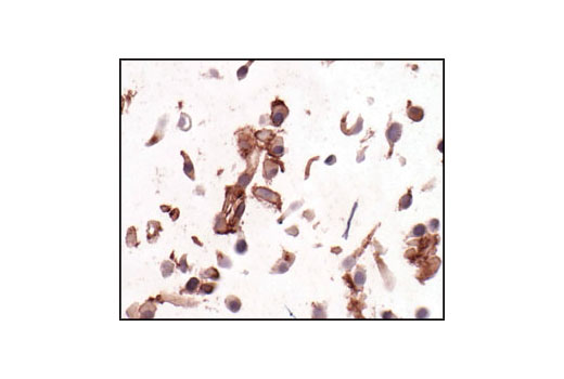
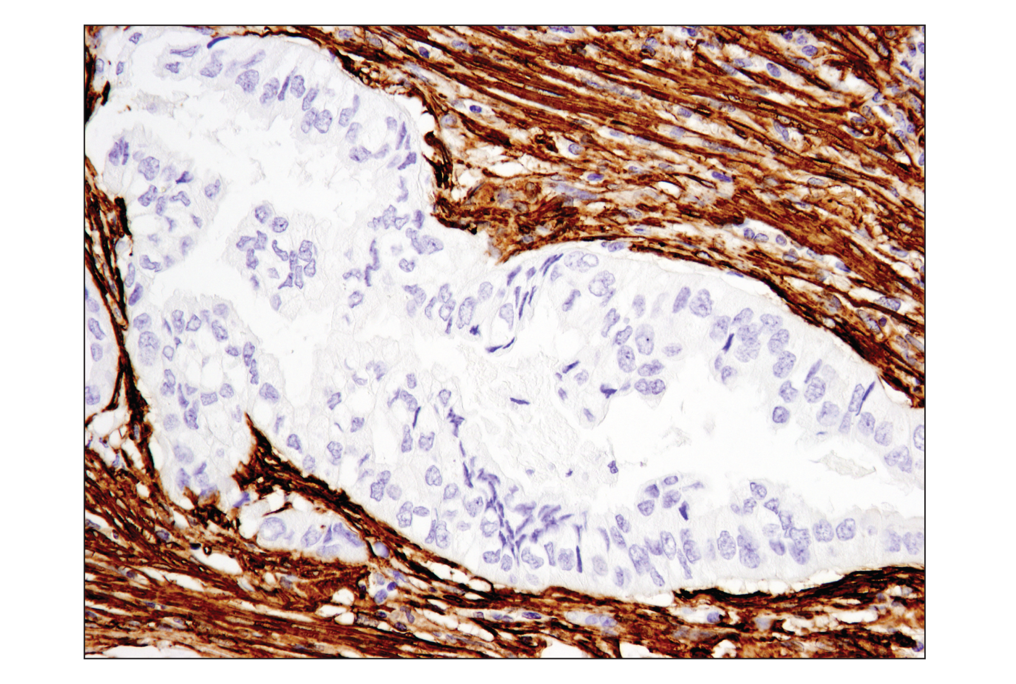
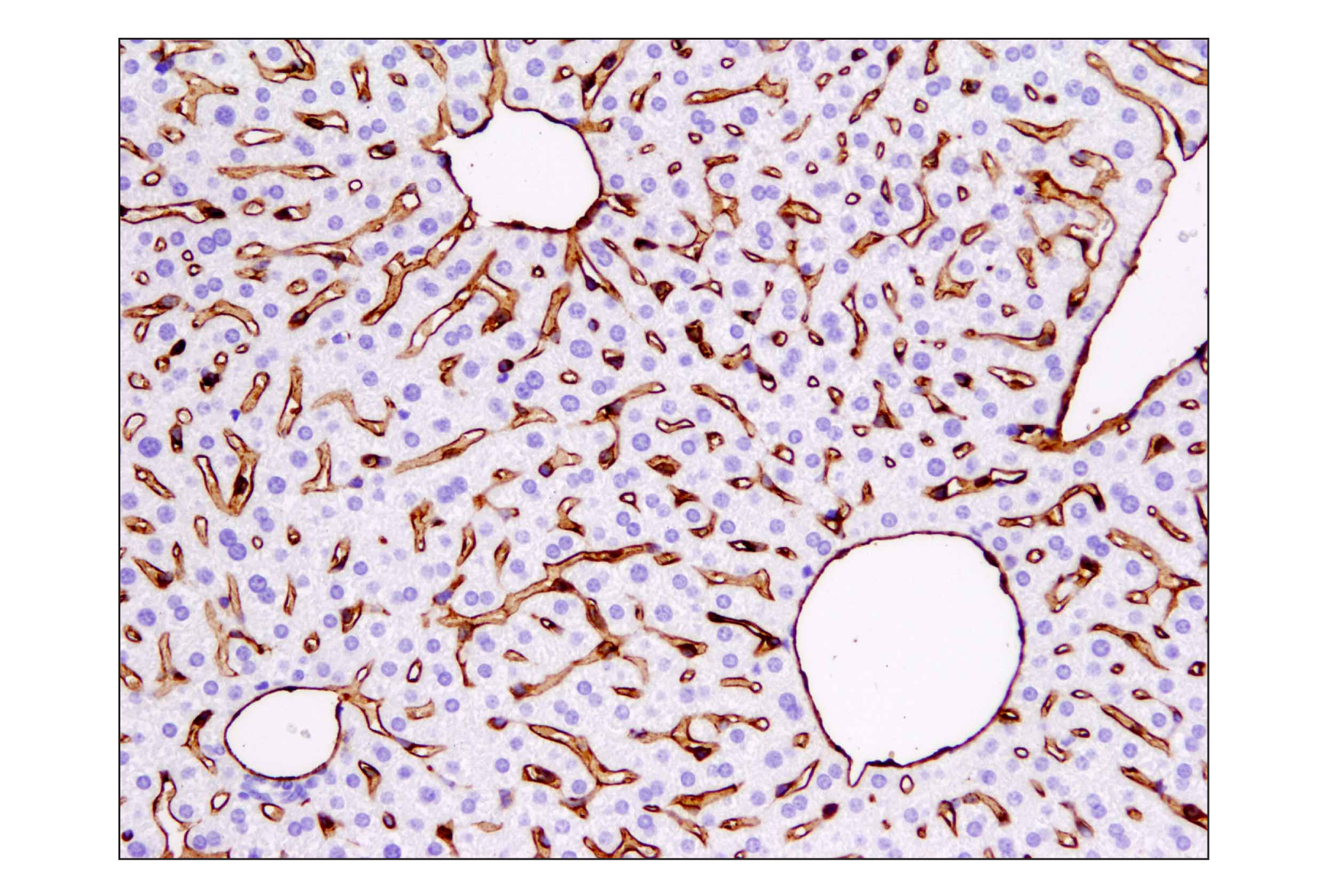
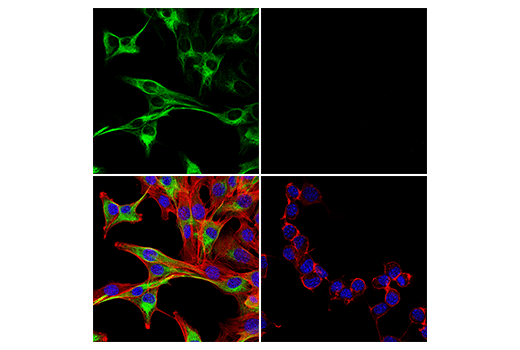
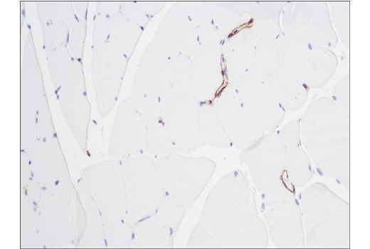
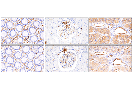
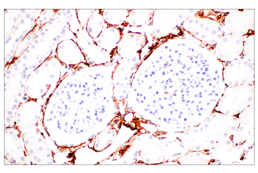
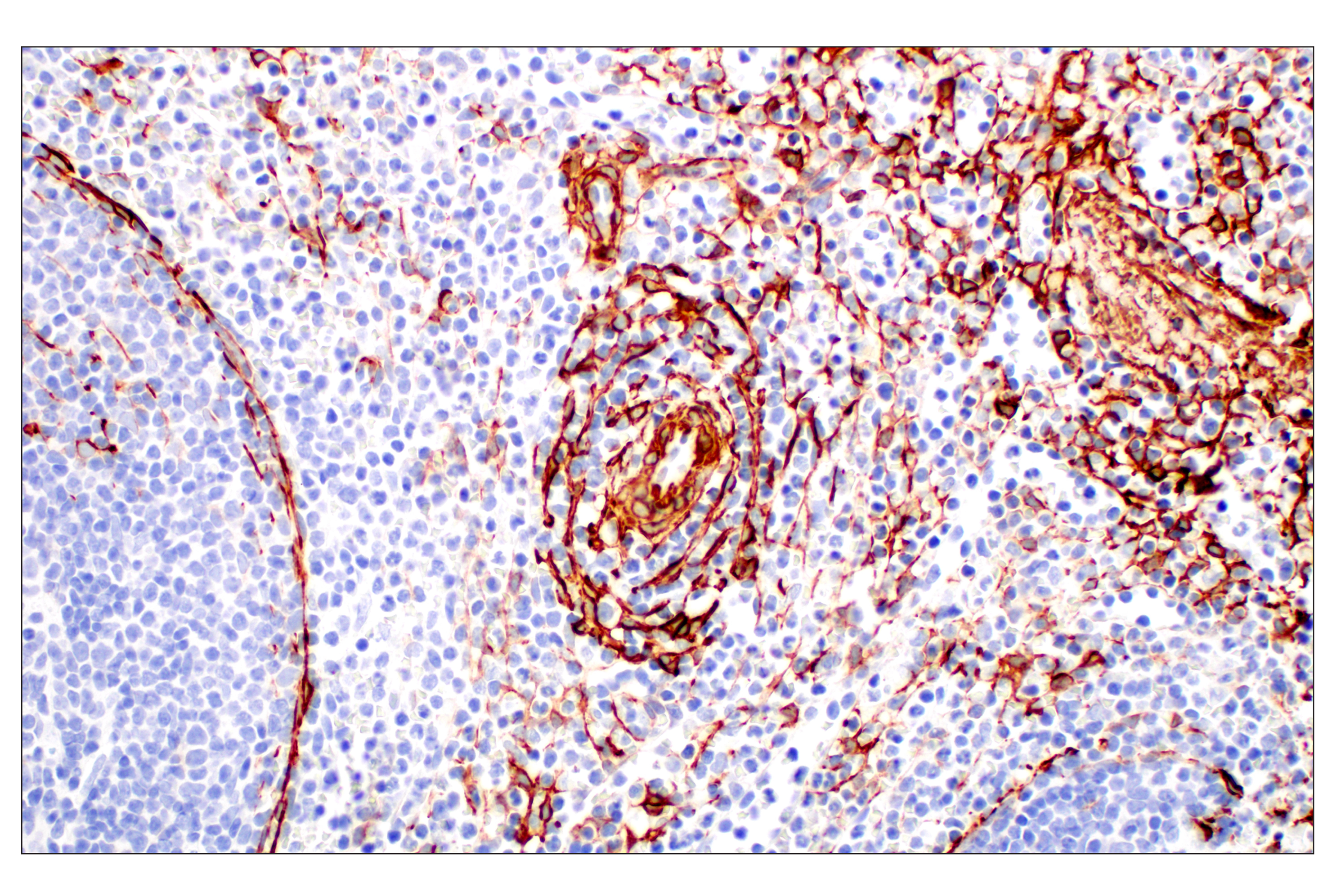
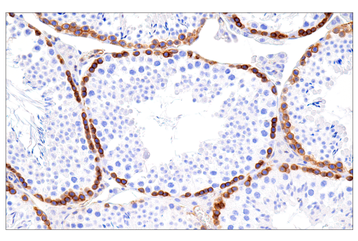
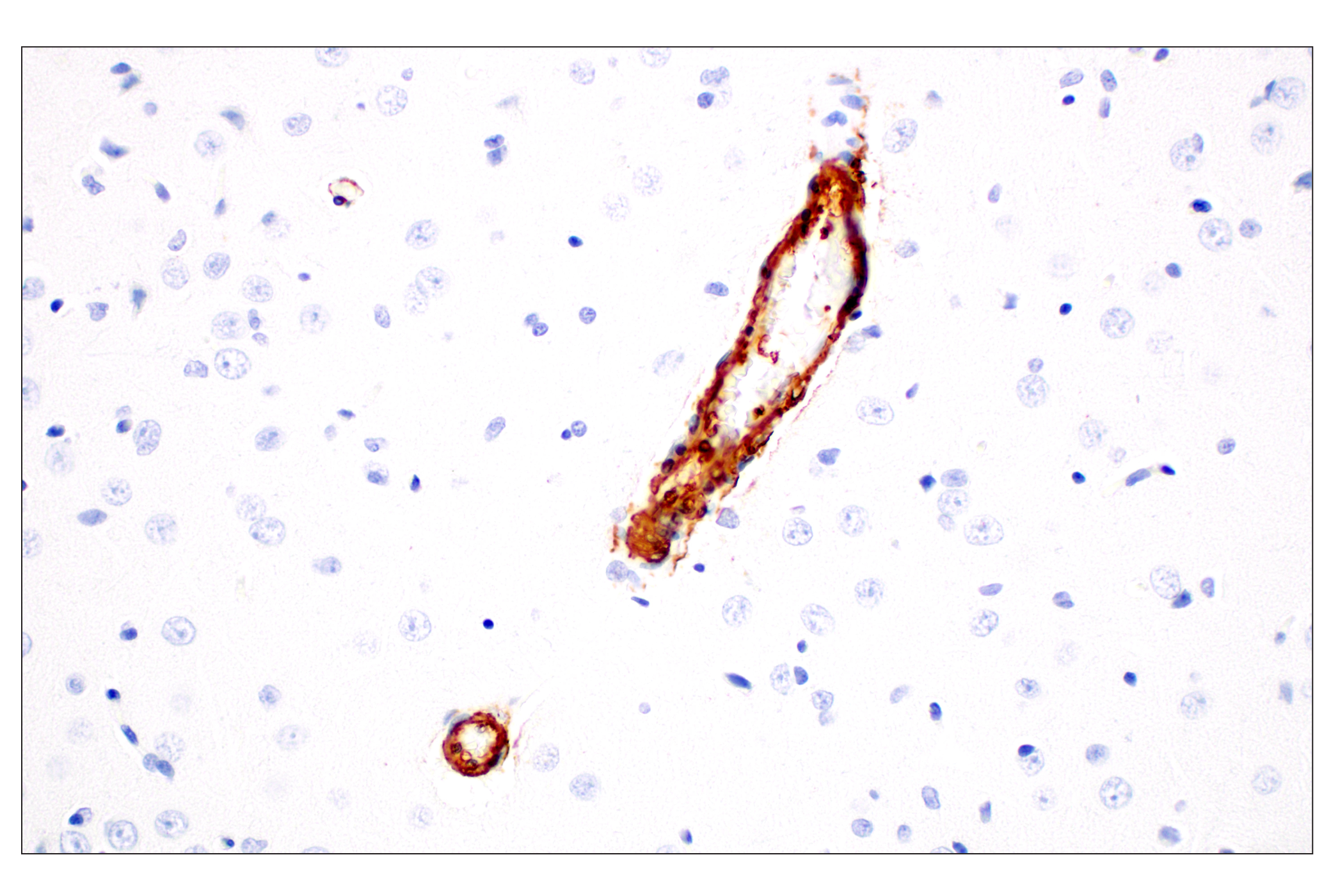
Immunohistochemical analysis of paraffin-embedded rat brain using alpha-Smooth Muscle Actin (D4K9N) XP® Rabbit mAb.
| Product Includes | Quantity | Applications | Reactivity | MW(kDa) | Isotype |
|---|---|---|---|---|---|
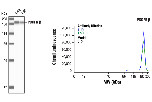
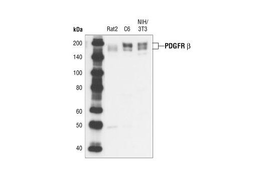
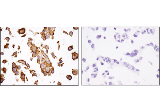
| PDGF Receptor β (28E1) Rabbit mAb 3169 | 20 µl |
|
H M R | 190 | Rabbit IgG |
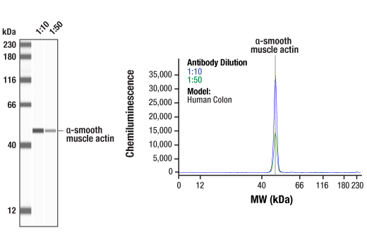
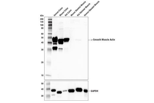
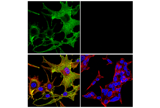
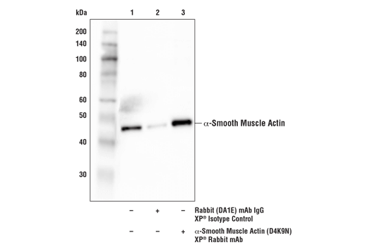
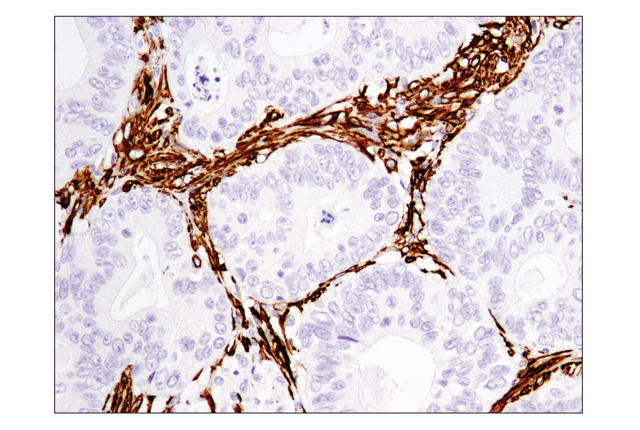
| α-Smooth Muscle Actin (D4K9N) XP® Rabbit mAb 19245 | 20 µl |
|
H M R Hm Mk | 42 | Rabbit IgG |
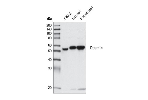
| Desmin (D93F5) XP® Rabbit mAb 5332 | 20 µl |
|
H M R | 53 | Rabbit IgG |
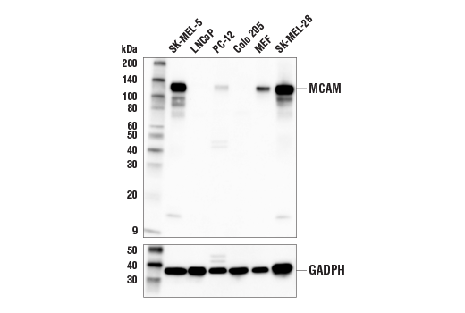
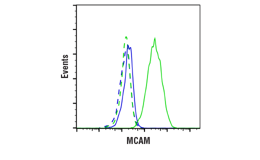
| MCAM (E3F3E) XP® Rabbit mAb 81701 | 20 µl |
|
H M | 120 | Rabbit IgG |
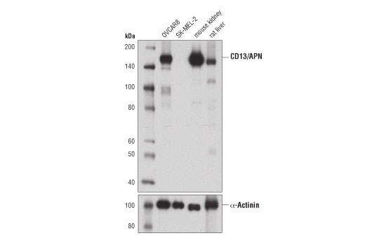
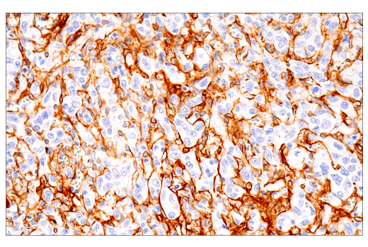
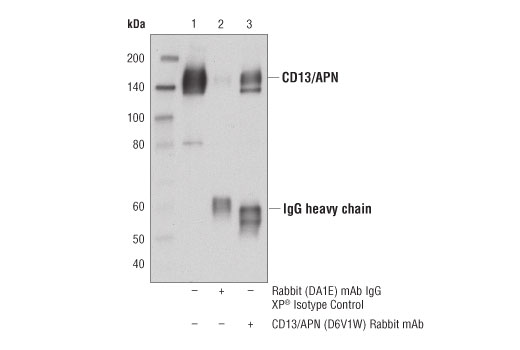
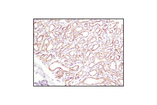
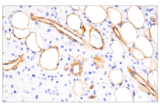
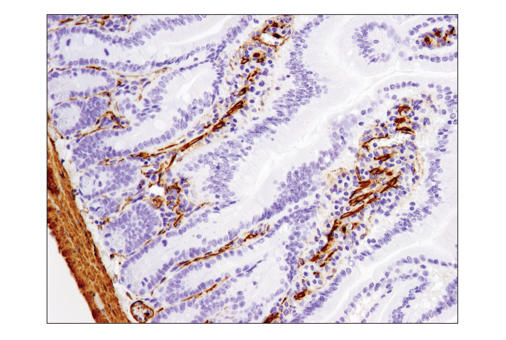
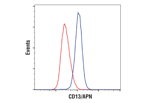
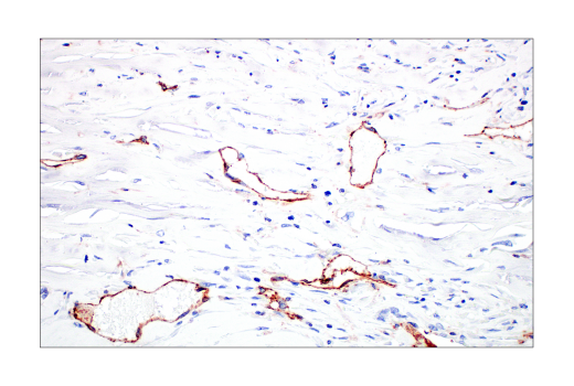
| CD13/APN (D6V1W) Rabbit mAb 32720 | 20 µl |
|
H M R | 160 | Rabbit IgG |
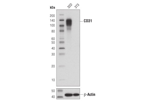
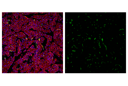
| CD31 (PECAM-1) (D8V9E) XP® Rabbit mAb 77699 | 20 µl |
|
M | 135 | Rabbit IgG |
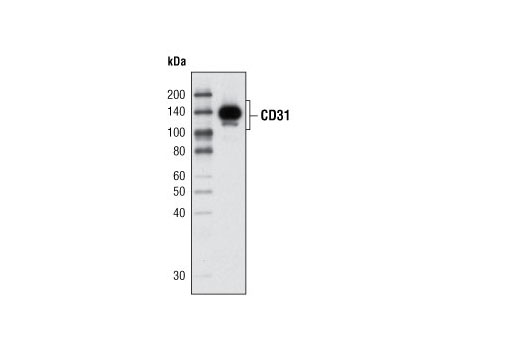
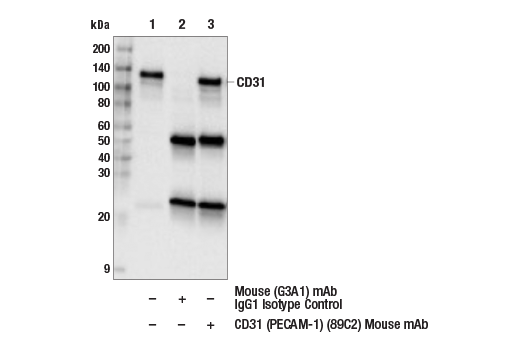
| CD31 (PECAM-1) (89C2) Mouse mAb 3528 | 20 µl |
|
H | 130 | Mouse IgG1 |
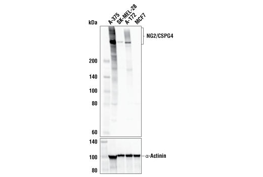
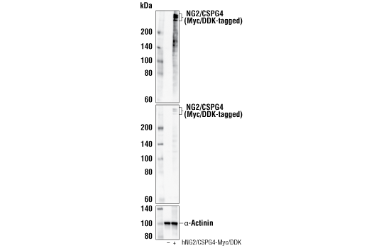
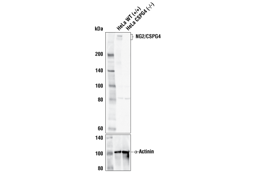
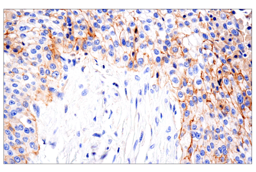
| NG2/CSPG4 (E3B3G) XP® Rabbit mAb 43916 | 20 µl |
|
H | 250, 450 | Rabbit IgG |
| Anti-rabbit IgG, HRP-linked Antibody 7074 | 100 µl |
|
Rab | Goat | |
| Anti-mouse IgG, HRP-linked Antibody 7076 | 100 µl |
|
M | Horse |
Product Information
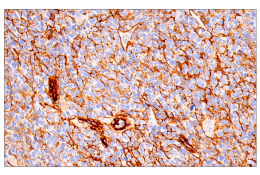
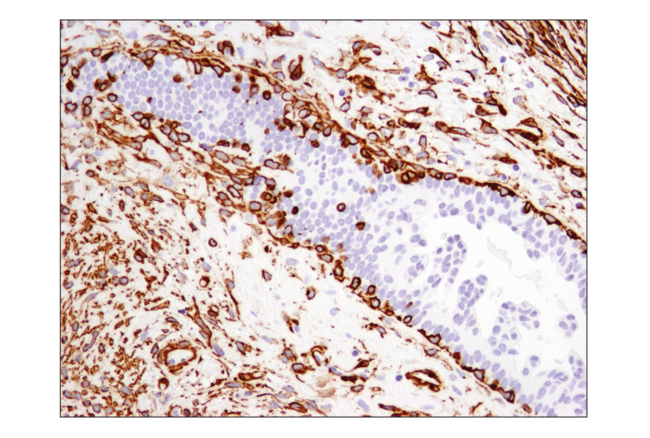
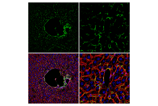
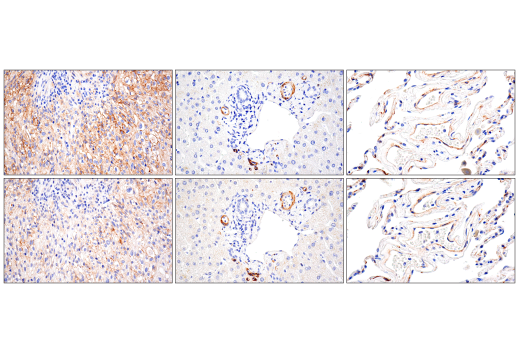
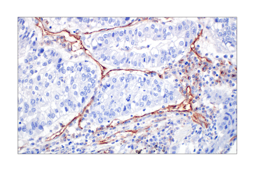
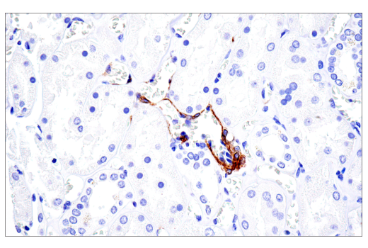
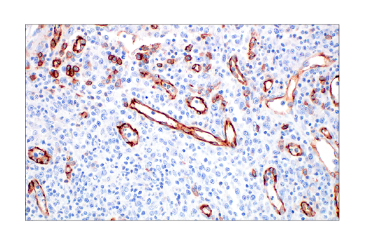
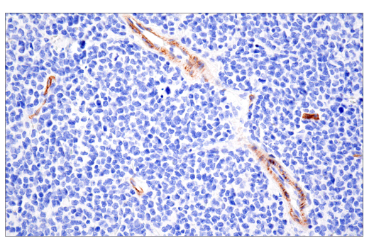
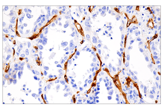
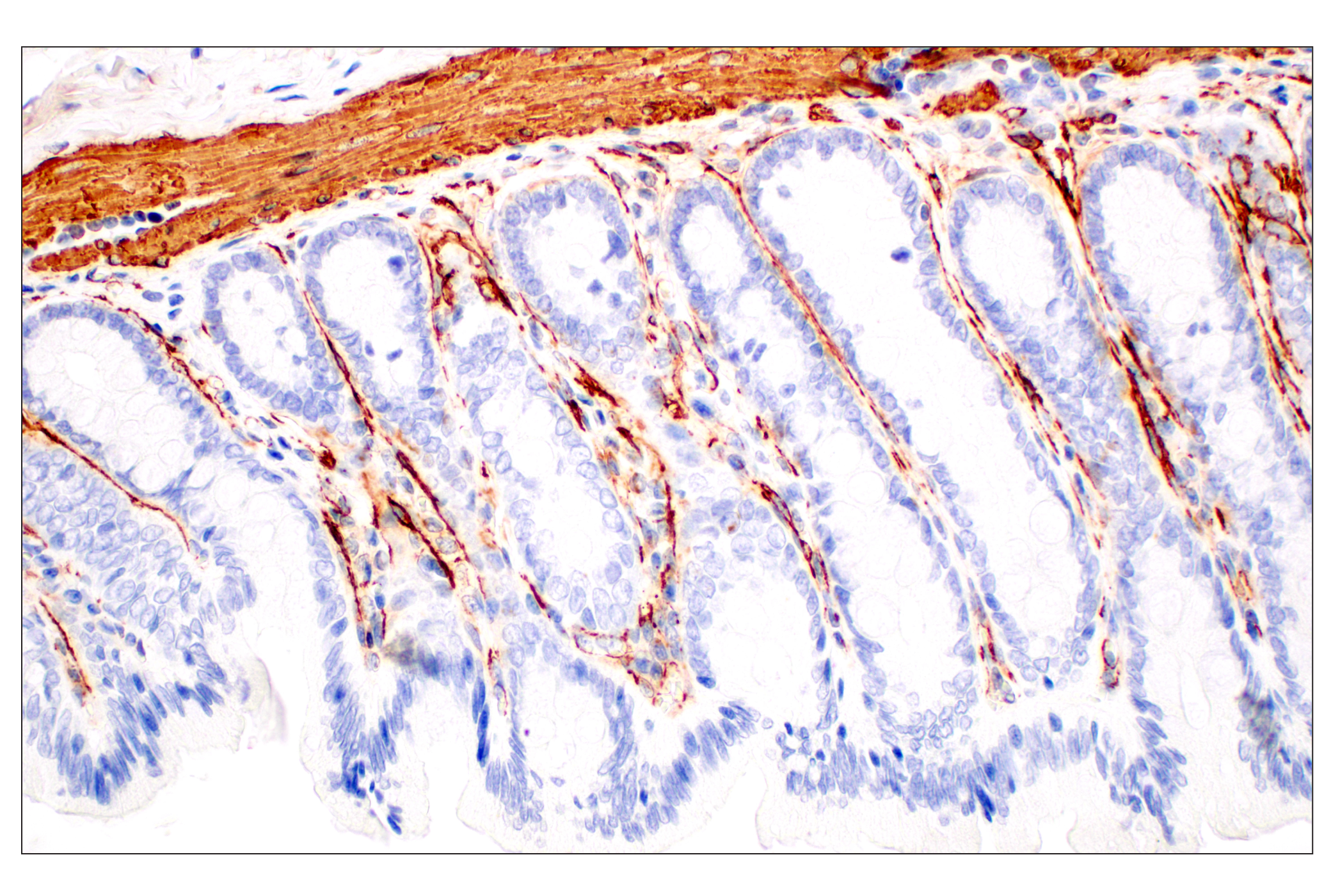
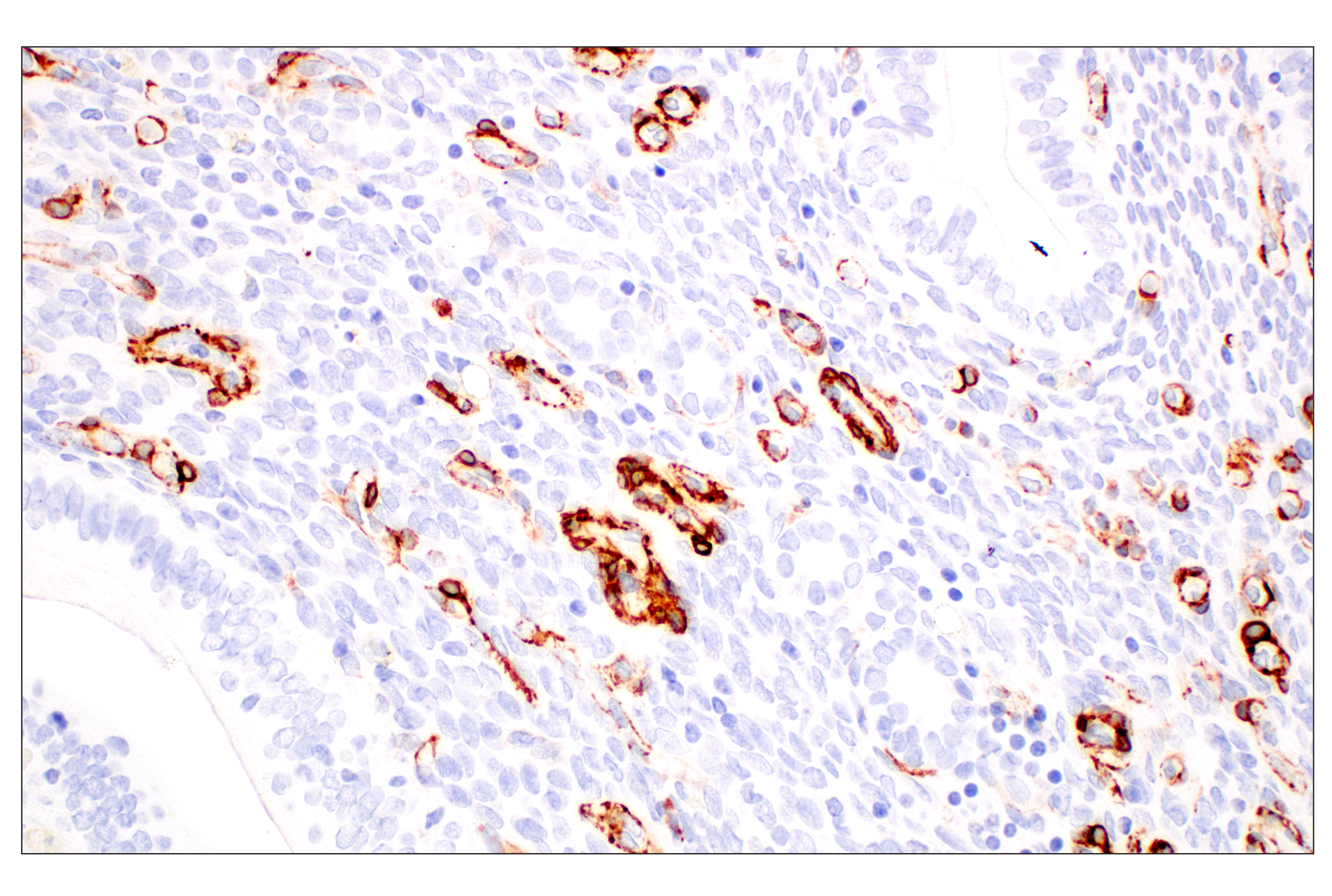
The antibodies in this kit serve to detect and evaluate blood-brain barrier (BBB) integrity in the brain. The BBB is formed by microvascular endothelial cells (ECs) lining the cerebral capillaries penetrating the brain and spinal cord of most mammals, providing a biological barrier at the blood to brain interface effectively separating the brain from the rest of the body (1). Pericytes are essential constituents of the brain capillary; their close association with ECs allows the exchange of ions, metabolites, second messengers, and ribonucleic acids between the two cell types (1). Pericyte markers include smooth muscle actin, desmin, or platelet-derived growth factor receptor β (PDGFR-β). Pericytes and ECs are in close communication with each other, for example, via the PDGF-β signaling pathway (2).
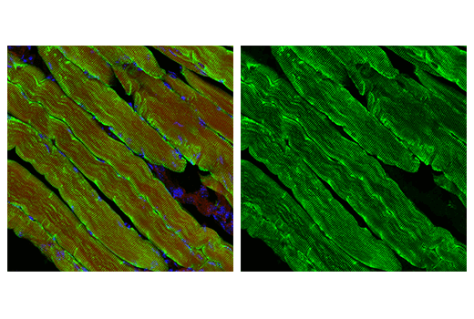
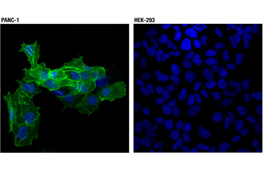
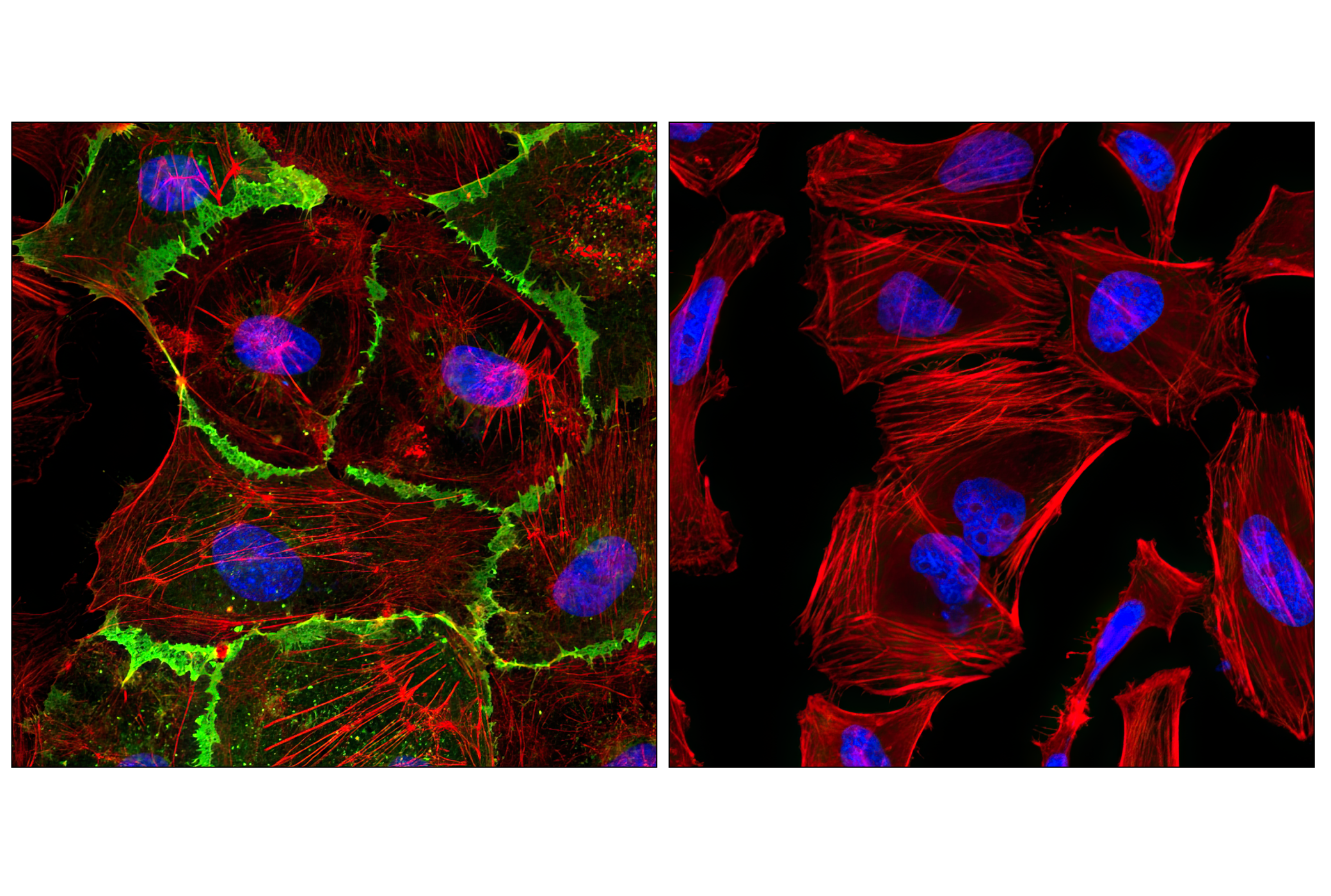
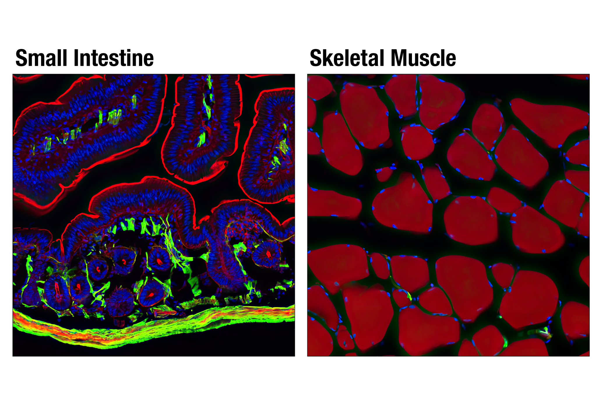
The cytoskeleton consists of three types of cytosolic fibers: microfilaments (actin filaments), intermediate filaments, and microtubules. Actin proteins are major components of the eukaryotic cytoskeleton. At least six vertebrate actin isoforms have been identified. The cytoplasmic β- and γ-actin proteins are referred to as “non-muscle” actin proteins as they are predominantly expressed in non-muscle cells where they control cell structure and motility (3). The α-cardiac and α-skeletal actin proteins are expressed in striated cardiac and skeletal muscles, respectively. The smooth muscle α-actin and γ-actin proteins are found primarily in vascular smooth muscle and enteric smooth muscle, respectively. The α-smooth muscle actin (ACTA2) is also known as aortic smooth muscle actin. These actin isoforms regulate the contractile potential of muscle cells (4).
The intermediate filament desmin is a myogenic marker expressed in early development that forms a network of filaments extending across the myofibril and surrounding Z discs. The desmin cytoskeleton provides a connection among myofibrils, organelles, and the cytoskeleton (5). Melanoma cell adhesion molecule (MCAM, MUC18, CD146) is a marker protein seen in vascular endothelial cells, activated T lymphocytes, smooth muscle, and bone marrow stromal cells. Endothelial MCAM within the BBB acts as adhesion receptors that permit lymphocytes to transmigrate across the barrier and produce inflammatory lesions (6). MCAM also functions as a co-receptor for PDGFR-β on pericytes to regulate pericyte-EC interactions (7).
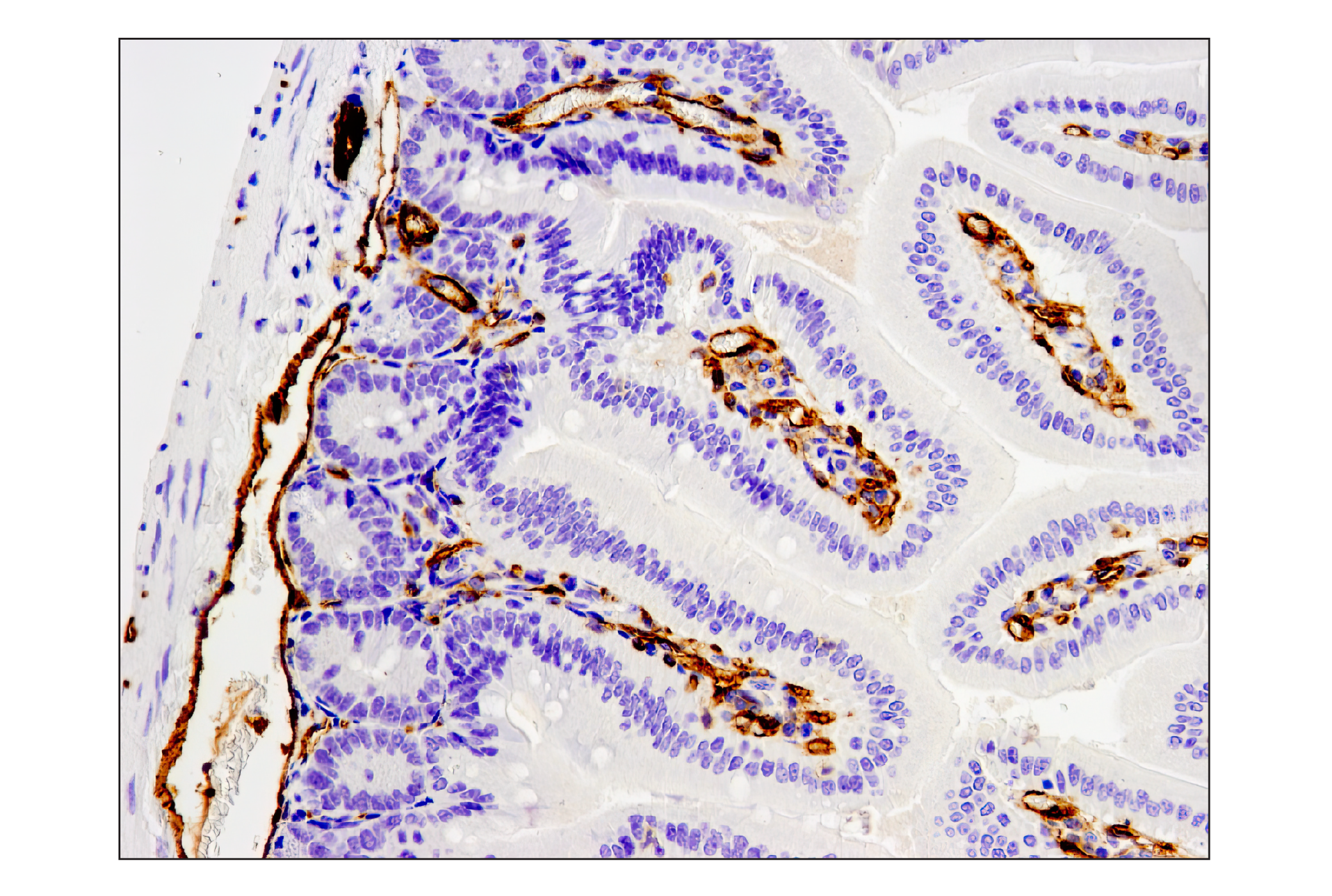
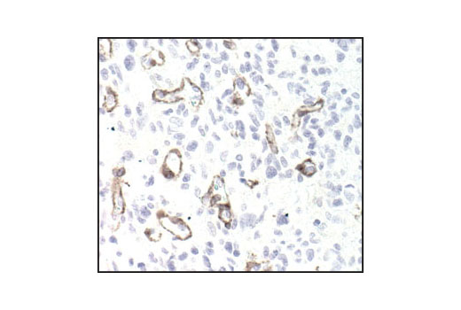
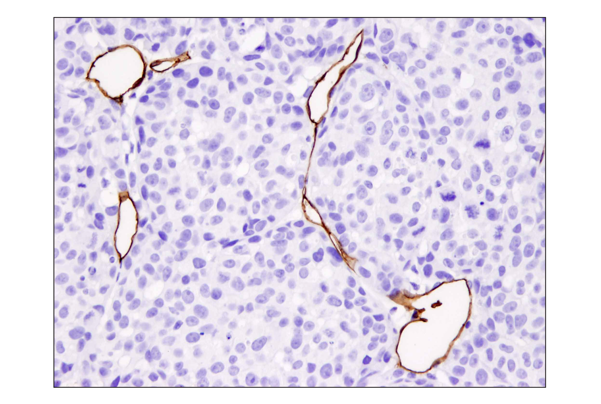
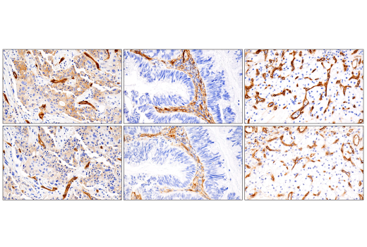
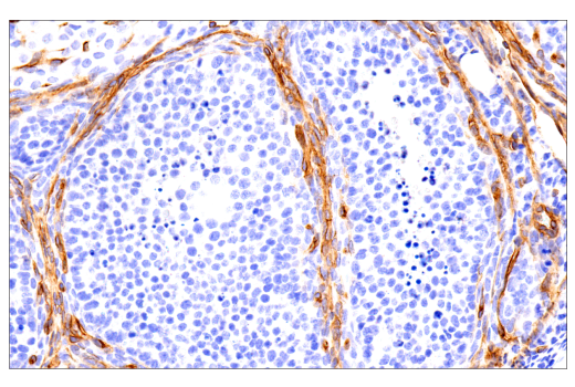
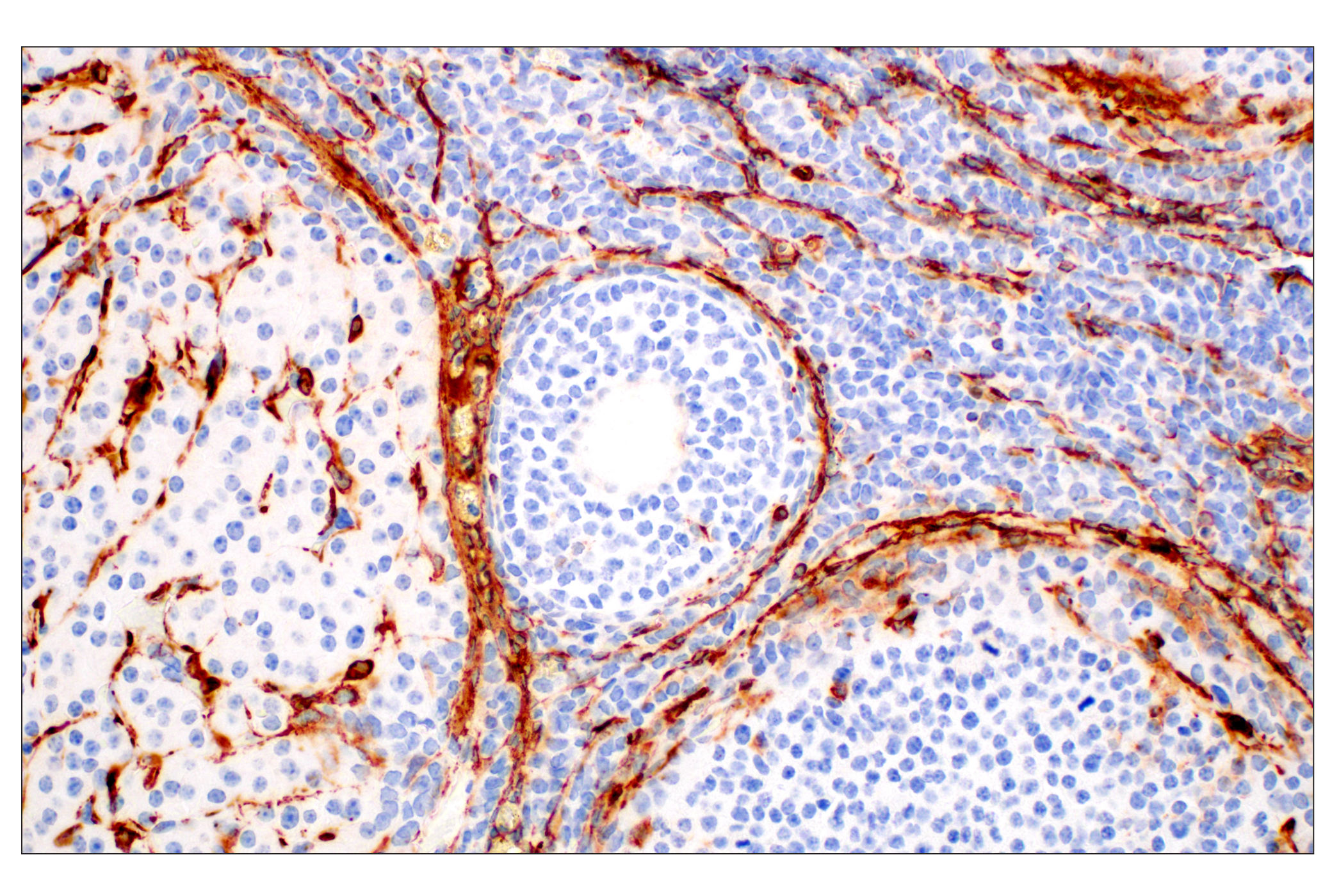
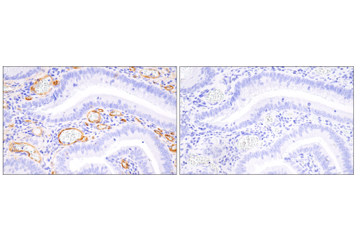
CD31 (Platelet Endothelial Cell Adhesion Molecule-1: PECAM-1), a member of the Ig superfamily of cell adhesion molecules, is expressed by circulating platelets, monocytes, neutrophils, some T cells, and endothelial cells and modulates cell adhesion, endothelial cell migration, and angiogenesis. CD31 serves as a scaffold for various signaling molecules and is a possible gate-keeper receptor in the inflammatory blood–brain axis (8).
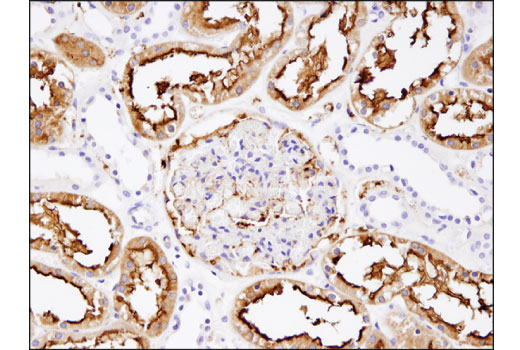
Aminopeptidase N (APN, CD13) is a metalloprotease expressed on myeloid cells, pericytes, fibroblasts, epithelial and endothelial cells, as well as on tumor cells, and stem cells. APN/CD13 acts as an adhesion molecule that modulates inflammatory immune cell trafficking, resulting in injury progression (9).
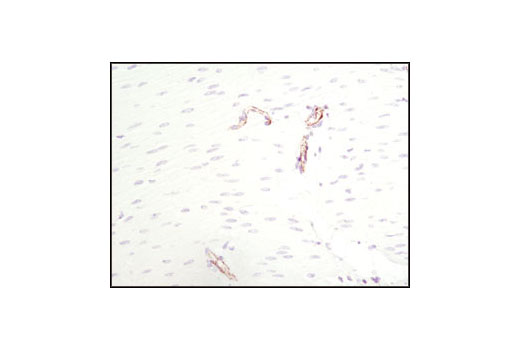
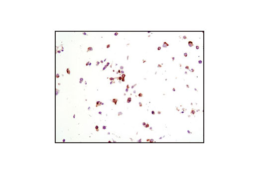
The chondroitin sulfate proteoglycan NG2 is a type I membrane protein expressed by subpopulations of glia, including oligodendroglial precursor cells and a variety of tumor cells (10). Although NG2 is not expressed in adult central nervous system (CNS) pericytes, it is an early marker of pericyte activation during CNS development and pathological conditions. Thus, upon CNS injury, NG2-reactive pericytes are found along microvessels, where they act as sensors for inflammation and support the immunosurveillance and effector function of extravasated neutrophils and macrophages (11).
Explore pathways related to this product.
STRING - Known and Predicted Protein-Protein Interactions.
Except as otherwise expressly agreed in a writing signed by a legally authorized representative of CST, the following terms apply to Products provided by CST, its affiliates or its distributors. Any Customer's terms and conditions that are in addition to, or different from, those contained herein, unless separately accepted in writing by a legally authorized representative of CST, are rejected and are of no force or effect.
Products are labeled with For Research Use Only or a similar labeling statement and have not been approved, cleared, or licensed by the FDA or other regulatory foreign or domestic entity, for any purpose. Customer shall not use any Product for any diagnostic or therapeutic purpose, or otherwise in any manner that conflicts with its labeling statement. Products sold or licensed by CST are provided for Customer as the end-user and solely for research and development uses. Any use of Product for diagnostic, prophylactic or therapeutic purposes, or any purchase of Product for resale (alone or as a component) or other commercial purpose, requires a separate license from CST. Customer shall (a) not sell, license, loan, donate or otherwise transfer or make available any Product to any third party, whether alone or in combination with other materials, or use the Products to manufacture any commercial products, (b) not copy, modify, reverse engineer, decompile, disassemble or otherwise attempt to discover the underlying structure or technology of the Products, or use the Products for the purpose of developing any products or services that would compete with CST products or services, (c) not alter or remove from the Products any trademarks, trade names, logos, patent or copyright notices or markings, (d) use the Products solely in accordance with CST Product Terms of Sale and any applicable documentation, and (e) comply with any license, terms of service or similar agreement with respect to any third party products or services used by Customer in connection with the Products.